. 2018 Nov 6;8(1):16394.
doi: 10.1038/s41598-018-34809-6.

Direct quantitative material decomposition employing grating-based X-ray phase-contrast CT

Affiliations

Direct quantitative material decomposition employing grating-based X-ray phase-contrast CT

Eva Braig et al. Sci Rep. .

Erratum in

Abstract

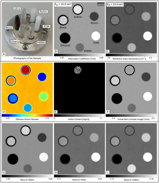
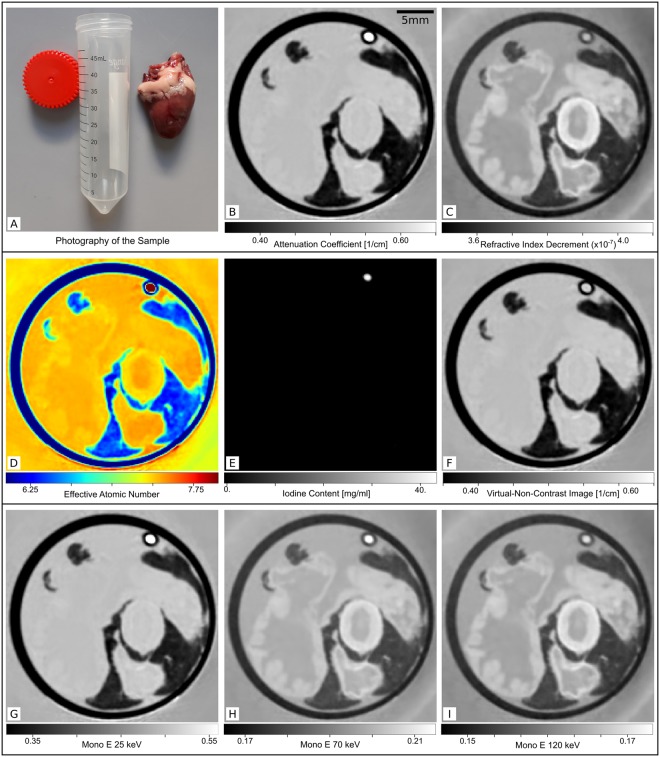
Dual-energy CT has opened up a new level of quantitative X-ray imaging for many diagnostic applications. The energy dependence of the X-ray attenuation is the key to quantitative material decomposition of the volume under investigation. This material decomposition allows the calculation of virtual native images in contrast enhanced angiography, virtual monoenergetic images for beam-hardening artifact reduction and quantitative material maps, among others. These visualizations have been proven beneficial for various diagnostic questions. Here, we demonstrate a new method of 'virtual dual-energy CT' employing grating-based phase-contrast for quantitative material decomposition. Analogue to the measurement at two different energies, the applied phase-contrast measurement approach yields dual information in form of a phase-shift and an attenuation image. Based on these two image channels, all known dual-energy applications can be demonstrated with our technique. While still in a preclinical state, the method features the important advantages of direct access to the electron density via the phase image, simultaneous availability of the conventional attenuation image at the full energy spectrum and therefore inherently registered image channels. The transfer of this signal extraction approach to phase-contrast data multiplies the diagnostic information gained within a single CT acquisition. The method is demonstrated with a phantom consisting of exemplary solid and fluid materials as well as a chicken heart with an iodine filled tube simulating a vessel. For this first demonstration all measurements have been conducted at a compact laser-undulator synchrotron X-ray source with a tunable X-ray energy and a narrow spectral bandwidth, to validate the quantitativeness of the processing approach.


Conflict of interest statement

The authors declare no competing interests.

Figures

Figure 1
Figure 1
Measurements and results of virtual dual-energy processing for a material phantom. (A) Photography of the sample consisting of 6 different solid or liquid materials. From the reconstructed attenuation (B) and phase-contrast (C) data an effective interaction energy of Eeffμ,δ=24.6,23.8keV was assigned via the literature value for the linear attenuation coefficient and the refractive index decrement of PMMA. The effective atomic number map (D) shows the distribution of Zeff ≈ 6.25 for Nylon to Zeff ≈ 7.99 for iodine. The quantitative iodine map (E) shows positive values of [I] ≈ 4.6 mg/ml only for the iodine solution. The virtual non-contrast image (F) is the conventional attenuation image where the identified iodine containing pixels are replaced with the attenuation value of water. The virtual monoenergetic image at E1VMI=25keV (G) looks very similar to the conventional attenuation image. For the higher energies (H,I), the contribution of the electron density increases and the virtual monoenergetic image at E3VMI=120keV looks very similar to the electron density image (which is simply proportional to the refractive index decrement image).
Figure 2
Figure 2
Measurements and results of virtual dual-energy processing for a biological soft tissue sample (chicken heart). (A) Photography of a fresh chicken heart next to the measurement container. The conventional attenuation image (B) shows very limited contrast only between the fatty tissue and the experimentally simulated iodine filled vessel. The phase-contrast image (C) reveals good contrast for the different anatomical structures like muscle, fat and blood vessels (most likely the aorta and two vessels of the low pressure system) but no contrast between contrast agent and the surrounding water. The effective atomic number map (D) reflects the situation of the conventional attenuation image with very low variations between the different structures besides fat and contrast agent. The quantitative iodine map (E) shows positive iodine concentrations of [I] ≈ 43 mg/ml only for the region of the contrast agent filled tube. The virtual non-contrast image (F) is the conventional attenuation image with the iodine containing pixels replaced with the attenuation value of water. At E1VMI=25keV, the virtual monoenergetic image (G) looks very similar to the conventional attenuation image. For the low atomic number soft tissue materials the Compton effect dominates the image formation already at E2VMI=70keV (H) and a difference to the virtual monoenergetic image at E3VMI=120keV (I) and the phase-contrast image is only visible for the iodine filled tube.

References

    1. Alvarez RE, Macovski A. Energy-selective reconstructions in X-ray computerised tomography. Phys. Medicine Biol. 1976;21:733. doi: 10.1088/0031-9155/21/5/002. - DOI - PubMed
    1. Silva AC, et al. Dual-energy (spectral) CT: Applications in abdominal imaging. RadioGraphics. 2011;31:1031–1046. doi: 10.1148/rg.314105159. - DOI - PubMed
    1. Naruto N, Itoh T, Noguchi K. Dual energy computed tomography for the head. Jpn. J. Radiol. 2017;36:69–80. doi: 10.1007/s11604-017-0701-4. - DOI - PubMed
    1. Rutherford RA, Pullan BR, Isherwood I. Measurement of effective atomic number and electron density using an EMI scanner. Neuroradiology. 1976;11:15–21. doi: 10.1007/BF00327253. - DOI - PubMed
    1. Genant HK, Boyd D. Quantitative bone mineral analysis using dual energy computed tomography. Investig. Radiol. 1977;12:545–551. doi: 10.1097/00004424-197711000-00015. - DOI - PubMed

Publication types