. 2018 Nov 6;8(1):16424.
doi: 10.1038/s41598-018-34700-4.

Emergence of a cholecystokinin/sulfakinin signalling system in Lophotrochozoa

Affiliations

Emergence of a cholecystokinin/sulfakinin signalling system in Lophotrochozoa

Julie Schwartz et al. Sci Rep. .

Abstract

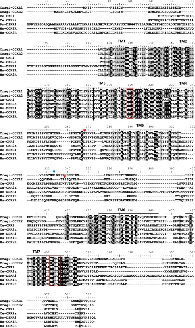
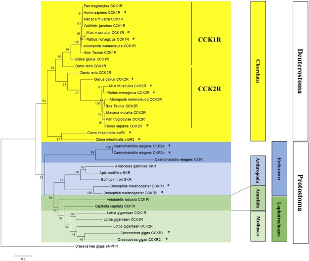
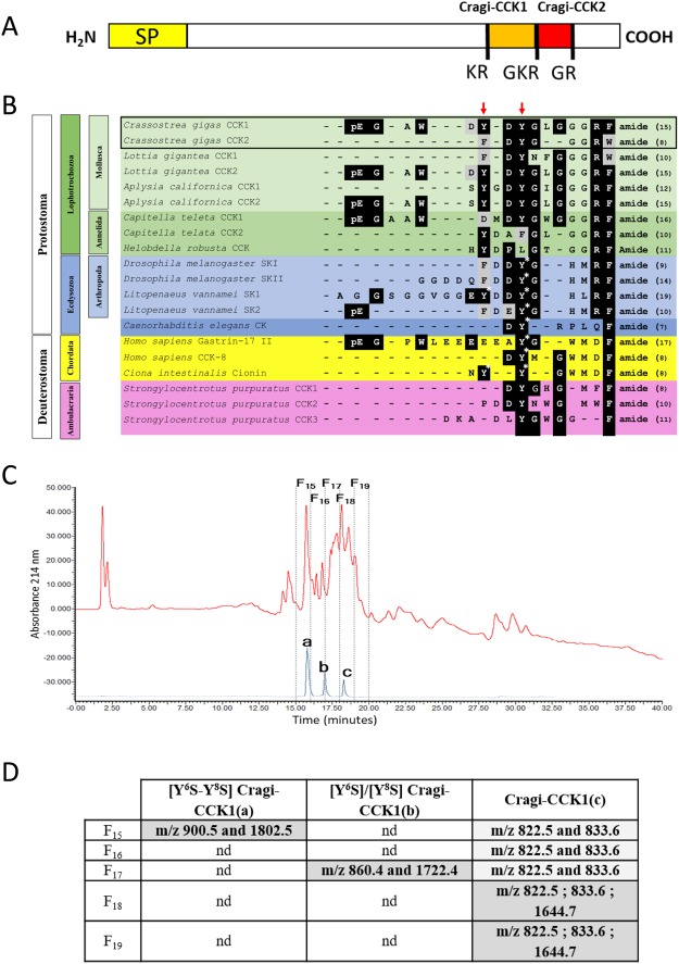
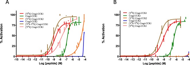
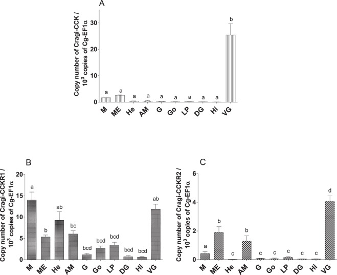
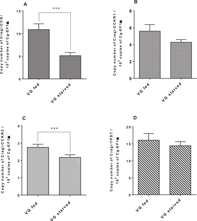
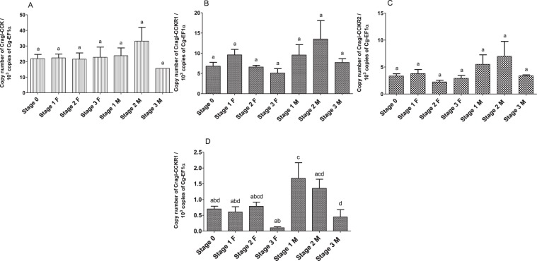
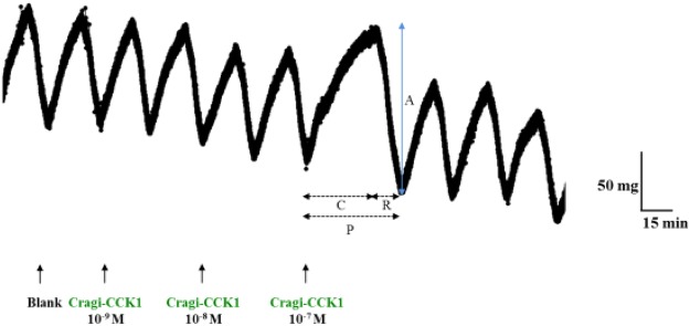
Chordate gastrin/cholecystokinin (G/CCK) and ecdysozoan sulfakinin (SK) signalling systems represent divergent evolutionary scenarios of a common ancestral signalling system. The present article investigates for the first time the evolution of the CCK/SK signalling system in a member of the Lophotrochozoa, the second clade of protostome animals. We identified two G protein-coupled receptors (GPCR) in the oyster Crassostrea gigas (Mollusca), phylogenetically related to chordate CCK receptors (CCKR) and to ecdysozoan sulfakinin receptors (SKR). These receptors, Cragi-CCKR1 and Cragi-CCKR2, were characterised functionally using a cell-based assay. We identified di- and mono-sulphated forms of oyster Cragi-CCK1 (pEGAWDY(SO3H)DY(SO3H)GLGGGRF-NH2) as the potent endogenous agonists for these receptors. The Cragi-CCK genes were expressed in the visceral ganglia of the nervous system. The Cragi-CCKR1 gene was expressed in a variety of tissues, while Cragi-CCKR2 gene expression was more restricted to nervous tissues. An in vitro bioassay revealed that different forms of Cragi-CCK1 decreased the frequency of the spontaneous contractions of oyster hindgut. Expression analyses in oysters with contrasted nutritional statuses or in the course of their reproductive cycle highlighted the plausible role of Cragi-CCK signalling in the regulation of feeding and its possible involvement in the coordination of nutrition and energy storage in the gonad. This study confirms the early origin of the CCK/SK signalling system from the common bilaterian ancestor and delivers new insights into its structural and functional evolution in the lophotrochozoan lineage.


Conflict of interest statement

The authors declare no competing interests.

Figures

Figure 1
Figure 1
Alignment of the amino acid sequence of Cragi-CCKR1 (MF787221), Cragi-CCKR2 (MF787222) with CCKR family members from Caenorhabditis elegans (Ce-CKR1: NP_491918.3; Ce-CKR2a: ACA81683.1), Drosophila melanogaster (Dm-DSKR1: NP_001097021.1), Homo sapiens (Hs-CCKR1: NP_000721.1; Hs-CCKR2: NP_795344.1) using CLUSTALW. Bars indicate the seven putative TM domains. Identical amino acid residues are highlighted in dark grey and similar residues in light grey. Black arrow indicates the position of a conserved intron, blue and red arrows indicate the respective position of Cragi-CCKR1 and Cragi-CCKR2 specific introns. Red boxes indicate the conserved “ERY” motif for receptor activation and “NPXXY” motif for Gαq and STAT –dependent signalling pathways. Red asterisk, the Arginine residue important for interaction with the sulphated tyrosine residue of CCK.
Figure 2
Figure 2
Phylogenetic representation of the relationship between Cragi-CCKRs and other CCKR family members. Phylogenetic and molecular evolutionary analyses were conducted using MEGA version 6 based on the maximum likelihood method. The accession members of the sequences used to construct the tree are listed in Supplementary Table 2. The Cg-sNPFR was chosen as outgroup. * indicates functionally characterized receptors.
Figure 3
Figure 3
(A) Schematic representation of Cragi-CCK precursor (SP: Signal peptide). (B) Sequence alignment of Cragi-CCK peptides with Deuterostome G/CCK family members, ecdysozoan sulfakinins (SK)/CK and lophotrochozoan CCK/SK,. The sulphated tyrosine residues (Y*) are labelled with an asterisk. Red-colored arrows indicate putative sulphated tyrosine residues in Cragi-CCKs or in putative CCK/SK peptides from other species. Black-colored and grey-colored shadings indicate respectively identical or similar amino acids (C) HPLC separation: In red, chromatogram of the second purification step of visceral ganglia extract. In blue, chromatogram of synthetic CCKs. a, [Y6S-Y8S] Cragi-CCK1; b, [Y6S]/[Y8S] Cragi-CCK1; c, Cragi-CCK1. (D) Detection with SIM mode of singly charged and doubled charged ions of different forms of Cragi-CCK1s in the different chromatographic fractions. nd: not detected.
Figure 4
Figure 4
Dose-dependent activity of Cragi-CCK peptides on Cragi-CCKR1 (A) and on Cragi-CCKR2 (B) expressed in HEK293T cells. Data are shown as relative (%) to the highest value (100% activation) for a given peptide and represent the mean of an experiment (n = 4) performed in triplicate. Vertical bars represent the standard error of the mean (SEM).
Figure 5
Figure 5
Distribution of mRNAs encoding Cragi-CCK (A), Cragi-CCKR1 (B) and Cragi-CCKR2 (C) in adult tissues. Each value is the mean + SEM of 5 pools of 6 animals. Expression levels were calculated as the number of copies of each specific transcript per 103 copies of Elongation Factor 1 α (EF1α) mRNA. Results were statistically tested with a one-way ANOVA p < 0,05. Significantly different means are indicated by different letters. M: Mantle; ME: Mantle Edge; He: heart; AM: Adductor Muscle; G: Gills; Go: Gonad; LP: Labial Palps; DG: Digestive Gland; Hi: Hindgut; VG: Visceral Ganglia.
Figure 6
Figure 6
Expression levels of Cragi-CCK (A), Cragi-CCKR1 (B), Cragi-CCKR2 (C), Cragi-TPST (D) mRNAs in VG of four weeks Isochrysis galbana fed or starved oysters. Each value is the mean + SEM of 15 independent animals (VG after conditioning with or without food). Results were statistically tested with a student’s t test. Significantly different means of samples from fed and starved animals are indicated by *(p < 0,05) (C) and ***(p < 0.001) (A). No significant statistical difference was observed for (B,D).
Figure 7
Figure 7
Level of expression of Cragi-CCK/Cragi-CCKR1/Cragi-CCKR2 mRNAs in VG (AC) and of Cragi-CCKR1 mRNA in the gonads (D) along an annual reproductive cycle. Each value is the mean + SEM of 5 pools of 6 animals. Results were statistically tested with a one-way ANOVA, p < 0,05. Samples with significant statistical difference are marked with distinct letters. F: Female; M: Male; 0: stage 0 (sexual resting stage); 1: stage 1 (gonial multiplication stage); 2: stage 2 (tubule development and maturation stage); 3: stage 3 (sexual maturity stage).
Figure 8
Figure 8
(A) Representative record of the hindgut-stimulating activity of Cragi-CCK1. Arrows indicate the application of saline solution (Blank) or Cragi-CCK1 peptide fractions at different concentrations. The different parameters taken into account to measure the biological response are indicated (C: Contraction time, R: Relaxation time, P: Period (min.) A: Amplitude of contraction (mg).

References

    1. Mirabeau O, Joly J. Molecular evolution of peptidergic signaling systems in bilaterians. Proc Natl Acad Sci USA. 2013;110:2028–2037. doi: 10.1073/pnas.1219956110. - DOI - PMC - PubMed
    1. Jékely G. Global view of the evolution and diversity of metazoan neuropeptide signaling. Proc Natl Acad Sci USA. 2013;110:8702–8707. doi: 10.1073/pnas.1221833110. - DOI - PMC - PubMed
    1. Cox KJ, et al. Cloning, characterization, and expression of a G-protein-coupled receptor from Lymnaea stagnalis and identification of a leucokinin-like peptide, PSFHSWSamide, as its endogenous ligand. J Neurosci. 1997;17:1197–1205. doi: 10.1523/JNEUROSCI.17-04-01197.1997. - DOI - PMC - PubMed
    1. Tensen CP, et al. The lymnaea cardioexcitatory peptide (LyCEP) receptor: a G-protein-coupled receptor for a novel member of the RFamide neuropeptide family. J Neurosci. 1998;18:9812–9821. doi: 10.1523/JNEUROSCI.18-23-09812.1998. - DOI - PMC - PubMed
    1. Moroz LL, et al. Neuronal transcriptome of Aplysia: neuronal compartments and circuitry. Cell. 2006;127:1453–67. doi: 10.1016/j.cell.2006.09.052. - DOI - PMC - PubMed

Publication types