. 2018 Nov 6;8(1):16423.
doi: 10.1038/s41598-018-34763-3.

Palm Fruit Bioactives modulate human astrocyte activity in vitro altering the cytokine secretome reducing levels of TNFα, RANTES and IP-10

Affiliations

Palm Fruit Bioactives modulate human astrocyte activity in vitro altering the cytokine secretome reducing levels of TNFα, RANTES and IP-10

Robert P Weinberg et al. Sci Rep. .

Abstract

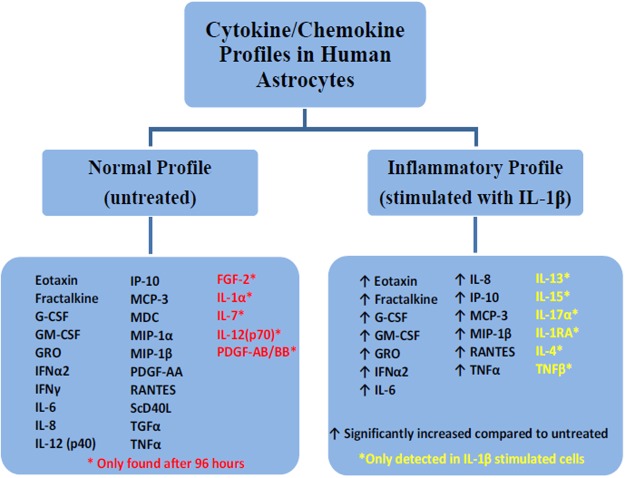
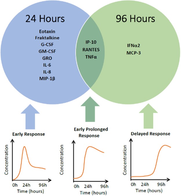
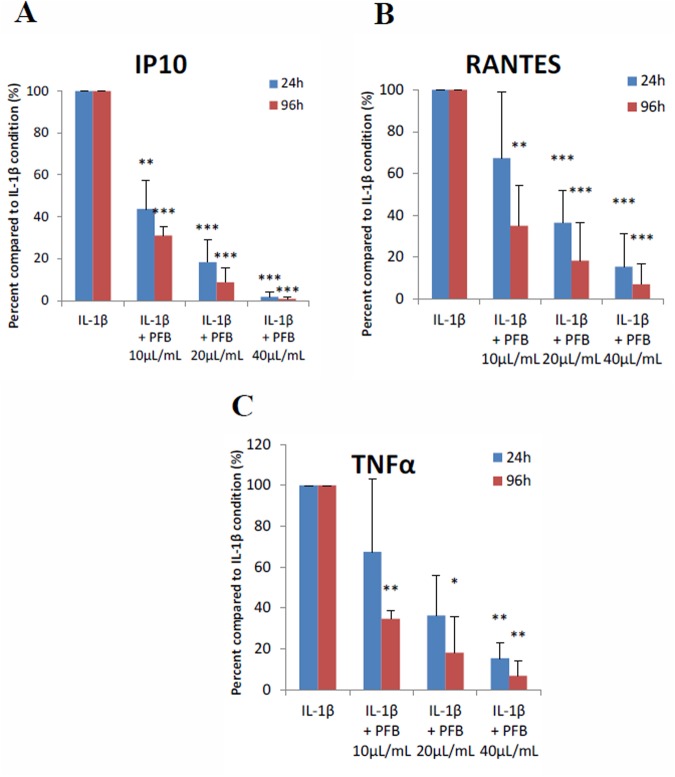
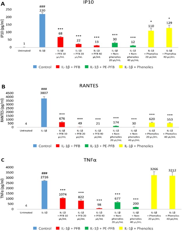
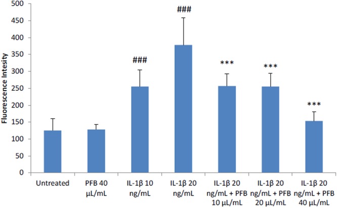
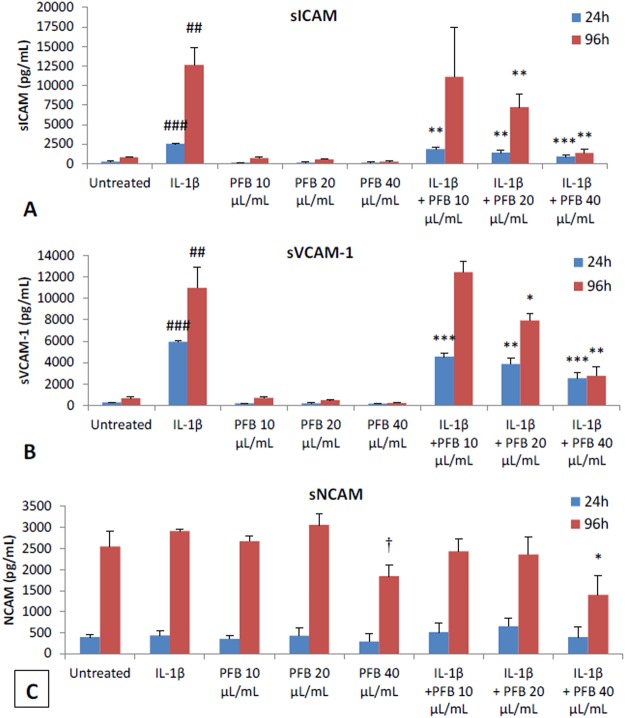
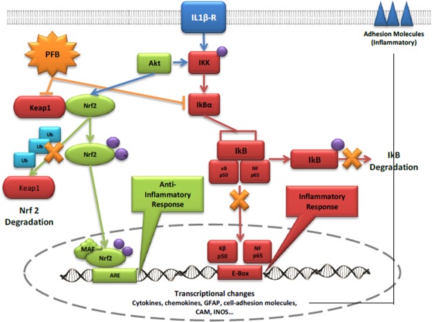
Neurodegenerative diseases, including Alzheimer's disease and Parkinson's disease, are becoming more prevalent and an increasing burden on society. Neurodegenerative diseases often arise in the milieu of neuro-inflammation of the brain. Reactive astrocytes are key regulators in the development of neuro-inflammation. This study describes the effects of Palm Fruit Bioactives (PFB) on the behavior of human astrocytes which have been activated by IL-1β. When activated, the astrocytes proliferate, release numerous cytokines/chemokines including TNFα, RANTES (CCL5), IP-10 (CXCL10), generate reactive oxygen species (ROS), and express specific cell surface biomarkers such as the Intercellular Adhesion Molecule (ICAM), Vascular Cellular Adhesion Molecule (VCAM) and the Neuronal Cellular Adhesion Molecule (NCAM). Interleukin 1-beta (IL-1β) causes activation of human astrocytes with marked upregulation of pro-inflammatory genes. We show significant inhibition of these pro-inflammatory processes when IL-1β-activated astrocytes are exposed to PFB. PFB causes a dose-dependent and time-dependent reduction in specific cytokines: TNFα, RANTES, and IP-10. We also show that PFB significantly reduces ROS production by IL-1β-activated astrocytes. Furthermore, PFB also reduces the expression of ICAM and VCAM, both in activated and naïve human astrocytes in vitro. Since reactive astrocytes play an essential role in the neuroinflammatory state preceding neurodegenerative diseases, this study suggests that PFB may have a potential role in their prevention and/or treatment.


Conflict of interest statement

Several of the authors have an equity interest in Phenolaeis, a nutraceutical company incorporated in Malaysia, which has not yet gone into production nor do any of the authors receive any royalties or profits from this entity.

Figures

Figure 1
Figure 1
Some bioactive organic compounds in Palm Fruit Bioactives (PFB). PFB is a complex heterogeneous phytochemical mixture prepared from the aqueous extract of the oil palm fruit.
Figure 2
Figure 2
Profiles of cytokines/chemokines produced by human astrocytes with/without IL-1β stimulation reveal 2 profiles: normal (basal) and inflammatory (post-IL-1β) The inflammatory profile consists of those secreted cytokines/chemokines which were significantly altered with IL-1β stimulation compared to their basal expression levels at 24 h or 96 h, while the normal profile consists of those cytokines/chemokines detectable in the basal state.
Figure 3
Figure 3
Time-dependent differential expression of cytokines/chemokines induced by IL-1β stimulation in human astrocytes. Cytokines induced within 24 hours are designated “early short response.” Cytokines produced after 96 hours of induction are designated “delayed response.” Cytokines induced within 24 h which remain elevated at 96 hours are designated “early-prolonged response”.
Figure 4
Figure 4
(A) IP-10, (B) RANTES, and (C) TNFα secretion levels induced in NHA by IL-1β stimulation (20 ng/ml) are significantly reduced with exposure to Palm Fruit Bioactives at 24 and 96 hours in a dose-dependent manner. The normalized percent change (%) for each PFB treatment condition compared with the positive control, IL-1β stimulation alone, for each time point. Data represent the mean percentage change ± SD of three Luminex replicates (*p < 0.05, **p < 0.01, ***p < 0.001).
Figure 5
Figure 5
Effects of phenolic vs. non-phenolic fractions of PFB on (A) IP10, (B) RANTES, and (C) TNFα levels in IL-1β-stimulated NHA. Concentrations of IP10, RANTES, TNFα for untreated and IL-1β stimulated controls, and IL-1β for different treatment conditions: PFB, non-phenolic PFB fraction, and phenolic PFB fraction at 20 and 40 μL/mL concentrations for each condition. Mean and standard deviation given for three replicates. Student’s t-test, ###p < 0.001, untreated vs. IL-1β stimulation; ***p < 0.001, IL-1β vs. IL-1β + PFB or PFB fraction.
Figure 6
Figure 6
Elevated Reactive Oxygen Species production in NHA stimulated by IL-1β is reduced by treatment with Palm Fruit Bioactives, as measured by DCFDA Assay after 24 h. No change is observed between ROS production between untreated NHA and the highest concentration of PFB treatment used, while increased ROS production from stimulation is decreased by PFB. Fluorescence intensity correlates with level of oxidative stress per condition in the DCDFA Assay. Values given as mean fluorescence intensity +/− standard deviation of three replicates. Student’s t-test, ###p < 0.001, untreated control vs. IL-1β; ***p < 0.001, IL-1β vs. PFB-X + IL-1β, where X is treatment concentration of PFB.
Figure 7
Figure 7
Effect of PFB on soluble adhesion molecules expressed by NHA with 24 h or 96 h stimulation with IL-1β. (A,B) PFB exposure decreased siCAM and sVCAM expression in both unstimulated and IL-1β-stimulated NHA. (C) NCAM expression did not differ upon exposure to IL-1β. Values given as cytokine concentration, mean +/− SD. Experiment was performed in triplicate. ##p < 0.01, ###p < 0.001, untreated control vs. IL-1β; *p < 0.05, **p < 0.01, ***p < 0.001, IL-1β vs. PFB-X + IL-1β.
Figure 8
Figure 8
Pro-inflammatory and anti-inflammatory pathways potentially affected by PFB. The pathways indicated by the red color represent the pro-inflammatory pathways mediated by NF-κB. The pathways indicated by the green color represent the anti-inflammatory pathways mediated by Nrf2.

Similar articles

Cited by

References

    1. Gordon MN, Schreier WA, Ou X, Holcomb LA, Morgan DG. Exaggerated astrocyte reactivity after nigrostriatal deafferentation in the aged rat. J Comp Neurol. 1997;388:106–19. doi: 10.1002/(SICI)1096-9861(19971110)388:1<106::AID-CNE7>3.0.CO;2-5. - DOI - PubMed
    1. Rozovsky I, Finch CE, Morgan TE. Age-related activation of microglia and astrocytes: In vitro studies show persistent phenotypes of aging, increased proliferation, and resistance to down-regulation. Neurobiol Aging. 1998;19:97–103. doi: 10.1016/S0197-4580(97)00169-3. - DOI - PubMed
    1. Apelt J, Leßig J, Schliebs R. β-Amyloid-associated expression of intercellular adhesion molecule-1 in brain cortical tissue of transgenic Tg2576 mice. Neurosci Lett. 2002;329:111–5. doi: 10.1016/S0304-3940(02)00589-X. - DOI - PubMed
    1. Griffin WS, Barger SW. Neuroinflammatory Cytokines-The Common Thread in Alzheimer’s Pathogenesis. US Neurol. 2010;6:19–27. doi: 10.17925/USN.2010.06.02.19. - DOI - PMC - PubMed
    1. Lee JK, Tran T, Tansey MG. Neuroinflammation in Parkinson’s disease. J Neuroimmune Pharmacol. 2009;4:419–29. doi: 10.1007/s11481-009-9176-0. - DOI - PMC - PubMed

Publication types

MeSH terms