. 2018 Nov 6;8(1):16437.
doi: 10.1038/s41598-018-34773-1.

Microinjection to deliver protein, mRNA, and DNA into zygotes of the cnidarian endosymbiosis model Aiptasia sp

Affiliations

Microinjection to deliver protein, mRNA, and DNA into zygotes of the cnidarian endosymbiosis model Aiptasia sp

Victor A S Jones et al. Sci Rep. .

Abstract

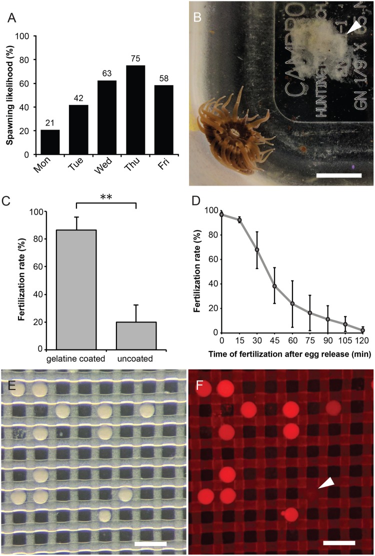
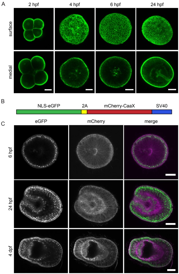
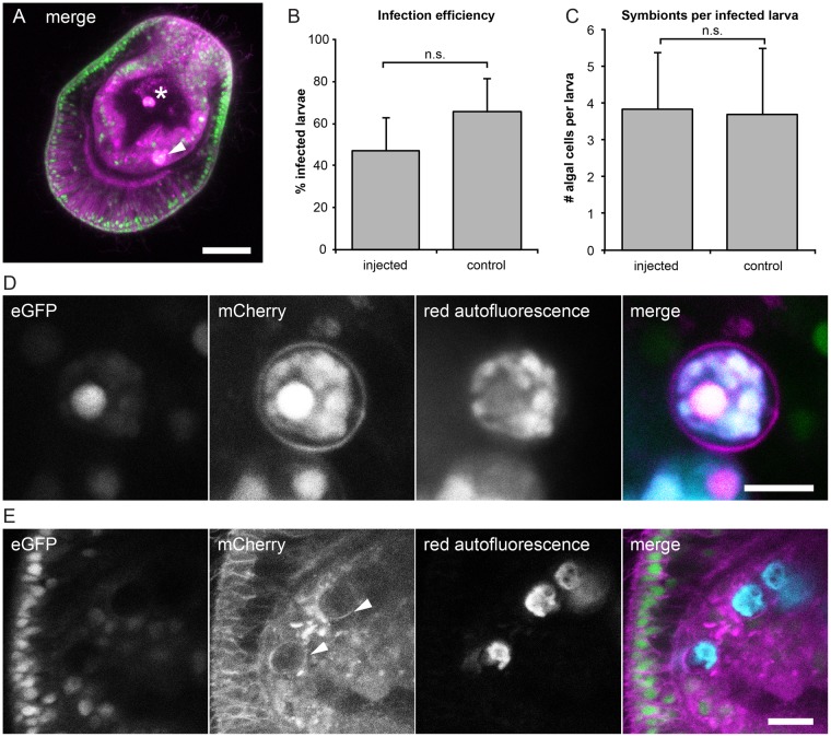
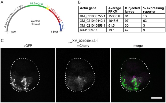
Reef-building corals depend on an intracellular symbiosis with photosynthetic dinoflagellates for their survival in nutrient-poor oceans. Symbionts are phagocytosed by coral larvae from the environment and transfer essential nutrients to their hosts. Aiptasia, a small tropical marine sea anemone, is emerging as a tractable model system for coral symbiosis; however, to date functional tools and genetic transformation are lacking. Here we have established an efficient workflow to collect Aiptasia eggs for in vitro fertilization and microinjection as the basis for experimental manipulations in the developing embryo and larvae. We demonstrate that protein, mRNA, and DNA can successfully be injected into live Aiptasia zygotes to label actin with recombinant Lifeact-eGFP protein; to label nuclei and cell membranes with NLS-eGFP and farnesylated mCherry translated from injected mRNA; and to transiently drive transgene expression from an Aiptasia-specific promoter, respectively, in embryos and larvae. These proof-of-concept approaches pave the way for future functional studies of development and symbiosis establishment in Aiptasia, a powerful model to unravel the molecular mechanisms underlying intracellular coral-algal symbiosis.


Conflict of interest statement

The authors declare no competing interests.

Figures

Figure 1
Figure 1
Spawning induction and in vitro fertilization in Aiptasia. (A) Culture and induction regime provides gametes throughout the week. Percentage of spawning events on each weekday (total from 24 weeks). (B) Female animal with egg patch (arrowhead). Scale = 5 mm. (C) Gelatin coating increases fertilization success. n = 3; p < 0.01; error bars, s.d. (D) Fertilization success rapidly decreases over time after spawning (n = 5). Error bars, s.d. (E) Zygotes are transferred into a microinjection dish filled with FASW, where they sink into holes in the mesh. Scale = 200 μm. (D) Co-injection of dextran-AlexaFluor 594 allows identification of successfully injected zygotes. Injected zygotes are brightly fluorescent whereas uninjected zygotes appear dark (arrowhead). Scale = 200 μm.
Figure 2
Figure 2
Microinjection of recombinant Lifeact-eGFP protein and mRNA to fluorescently label nuclei and cell outlines. (A) Zygotes injected with ~3.4 mg/ml Lifeact-eGFP protein were fixed 2, 4, 6 and 24 hpf. Maximum projections of 5 z-planes near the surface or centre of the embryo are shown. Scale = 25 μm. (B) Schematic representation of bicistronic in vitro transcribed NLS-eGFP-V2A-mCherry-CaaX mRNA: eGFP with a nuclear localization signal (NLS) is coupled to mCherry with a C-terminal CaaX box for farnesylation and insertion into the membrane. The fluorophores are separated by the self-cleaving V2A peptide. (C) mRNA-injected embryos were fixed 6 or 24 hpf or 4 days post-fertilization (dpf). Left panels show eGFP-labeled nuclei, middle panels show mCherry-labeled membranes, and the right panel shows the merged images (eGFP green; mCherry magenta). Scale = 25 μm.
Figure 3
Figure 3
Symbiosis establishment and live imaging in microinjected larvae. (A) Larvae expressing the injected NLS-eGFP-V2A-mCherry-CaaX mRNA and containing acquired symbiont cells (brightly autofluorescent in magenta mCherry channel). Symbionts within endodermal tissue (arrowhead) can be distinguished from those in the gastric cavity (asterisk). Larvae were incubated with Symbiodinium strain SSB01 at a concentration of 100,000 cells per ml for 3 days. Scale = 25 μm. (B) Infection efficiencies and (C) average number of algae per larva do not significantly differ in mRNA injected and control larvae. (D) Symbionts autofluoresce in the mCherry channel (magenta) and the red channel (cyan). The mCherry-labeled symbiosome membrane can be clearly seen surrounding the internalized symbiont. Larvae were injected with NLS-eGFP-2A-mCherry-CaaX mRNA and incubated for 1 day with Symbiodinium strain SSB01, before fixation at 3 dpf. Scale = 5 μm. (E) Symbionts and symbiosome membranes (arrowheads) can be imaged in vivo. Larva injected with NLS-eGFP-2A-mCherry-CaaX mRNA and incubated for 1 day with Symbiodinium strain SSB01, before embedding in agarose and imaging at 3 dpf. Scale = 10 μm.
Figure 4
Figure 4
Microinjection of DNA into Aiptasia zygotes. (A) Schematic of injected plasmids: the promoter of an Aiptasia actin gene drives expression of the NLS-eGFP-V2A-mCherry-CaaX reporter with a SV40 termination sequence. Meganuclease I-SceI recognition sites are indicated. (B) The promoter of actin gene XM_021049442.1 drives reporter expression in the majority of larvae coinjected with plasmid and the meganuclease I-SceI. For each promoter tested, the expression level (FPKM) (larvae transcriptomes), the number of larvae injected and analyzed, and the percentage of larvae in which GFP signal could be detected are given. (C) Coinjection of I-SceI and the prom XM_021049442.1: NLS-eGFP-V2A-mCherry-CAAX:SV40 plasmid causes strong, mosaic expression of the transgene, labeling nuclei (green) and membranes (magenta). 10 hpf, scale = 25 μm.

References

    1. Muscatine, L. The role of symbiotic algae in carbon and energy flux in coral reefs. In Coral Reefs (ed. Zubinsky, Z.) 75–87 (Elsevier, 1990).
    1. Yellowlees D, Rees TAV, Leggat W. Metabolic interactions between algal symbionts and invertebrate hosts. Plant Cell Environ. 2008;31:679–694. doi: 10.1111/j.1365-3040.2008.01802.x. - DOI - PubMed
    1. van Oppen M. In vitro establishment of symbiosis in Acropora millepora planulae. Coral Reefs. 2001;20:200. doi: 10.1007/s003380100167. - DOI
    1. Wakefield TS, Kempf SC. Development of host- and symbiont-specific monoclonal antibodies and confirmation of the origin of the symbiosome membrane in a cnidarian-dinoflagellate symbiosis. Biol. Bull. 2001;200:127–143. doi: 10.2307/1543306. - DOI - PubMed
    1. Rodriguez-Lanetty M, Wood-Charlson E, Hollingsworth L, Krupp D, Weis V. Temporal and spatial infection dynamics indicate recognition events in the early hours of a dinoflagellate/coral symbiosis. Mar. Biol. 2006;149:713–719. doi: 10.1007/s00227-006-0272-x. - DOI

Publication types