Review
. 2018 Oct;35(4):215-220.
doi: 10.1055/s-0038-1669467. Epub 2018 Nov 5.

Bone and Soft-Tissue Biopsies: What You Need to Know

Affiliations
Review

Bone and Soft-Tissue Biopsies: What You Need to Know

Dimitrios K Filippiadis et al. Semin Intervent Radiol. 2018 Oct.

Abstract

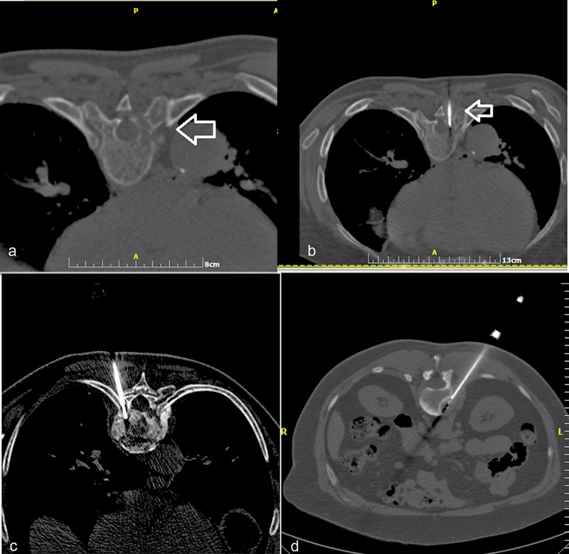
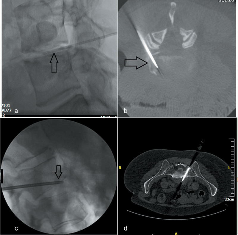
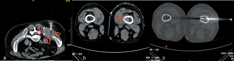
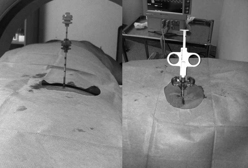
Percutaneous, image-guided musculoskeletal biopsy, due to its minimal invasive nature, when compared with open surgical biopsy, is a safe and effective technique which is widely used in many institutions as the primary method to acquire tissue and bone samples. Indications include histopathologic and molecular assessment of a musculoskeletal lesion, exclusion of malignancy in a bone/vertebral fracture, examination of bone marrow, and infection investigation. Preprocedural workup should include both imaging (for lesion assessment and staging) and laboratory (including coagulation tests and platelet count) studies. In selected cases, antibiotic prophylaxis should be administered before the biopsy. Core needle biopsy of musculoskeletal lesions has a diagnostic accuracy that ranges from 66 to 98% with higher diagnostic yield for lytic, large-size, malignant lesions and when multiple and long specimens are obtained. Reported complication rates range between 0 and 10% and usually do not exceed 5%, with a suggested threshold of 2%. The purpose of this review article is to illustrate the technical aspects, the indications, and the methodology of percutaneous image-guided bone biopsy that will assist the interventional radiologist to perform these minimal invasive techniques.

Keywords: Imaging guidance; interventional radiology; musculoskeletal lesion; percutaneous biopsy.


Conflict of interest statement

Conflict of Interest Authors have no conflict of interest to disclose.

Figures

Fig. 1
Fig. 1
( a ) Computed tomography (CT) axial scan illustrating a lytic lesion in the right pedicle of T8 vertebral body (arrow). ( b ) CT axial scan (same patient)—biopsy needle is placed inside the lesion (transpedicular approach) (arrow). ( c ) CT axial scan (different patient from a, b )—biopsy needle is placed inside T9 vertebral body (costovertebral approach). ( d ) CT axial scan (different patient from a, b , c )—biopsy needle is placed inside the L1 lesion (posterolateral approach).
Fig. 2
Fig. 2
Percutaneous biopsy for spondylodiscitis. ( a ) Lateral fluoroscopic view—needle is inside L4–L5 intervertebral disc (arrow)—biopsy is performed for culture in a patient with suspected spondylodiscitis. ( b ) Cone beam CT axial reconstruction (same patient)—needle is inside L4–L5 intervertebral disc (posterolateral approach). ( c ) Lateral fluoroscopic view (different patient from a, b )—biopsy needle (arrow) is placed inside L2–L3 intervertebral disc trough a transpedicular access crossing the end plate. Sampling is performed both from the end plate and the intervertebral disc. ( d ) Computed tomography axial scan (different patient from a, b, c )—biopsy needle is placed inside the lesion located anterior to the L5–S1 intervertebral disc (transsacral approach).
Fig. 3
Fig. 3
( a ) Computed tomography (CT) axial scan illustrating a large-size soft-tissue mass of the right iliac bone (arrows)—soft-tissue semiautomatic biopsy needle is placed in the periphery of the mass. ( b ) CT axial scan illustrating a hyperdense intraosseous lesion of the femoral bone. ( c ) CT axial scan (same patient with b )—after consulting with the surgeon, a lateral approach was performed; using a coaxial system, needle is located inside the lesion.
Fig. 4
Fig. 4
Coaxial approach offers the advantage of multiple bone sampling with a single puncture. Through the initial trocar, either a bone or a soft-tissue biopsy needle can be inserted depending on the lesion.

References

    1. Veltri A, Bargellini I, Giorgi L, Almeida P AMS, Akhan O. CIRSE guidelines on percutaneous needle biopsy (PNB) Cardiovasc Intervent Radiol. 2017;40(10):1501–1513. - PubMed
    1. Gogna A, Peh W C, Munk P L.Image-guided musculoskeletal biopsy Radiol Clin North Am 20084603455–473., v - PubMed
    1. Liu B, Limback J, Kendall M et al.Safety of CT-guided bone marrow biopsy in thrombocytopenic patients: a retrospective review. J Vasc Interv Radiol. 2017;28(12):1727–1731. - PubMed
    1. Holmes M G, Foss E, Joseph G et al.CT-guided bone biopsies in metastatic castration-resistant prostate cancer: factors predictive of maximum tumor yield. J Vasc Interv Radiol. 2017;28(08):1073–10810. - PubMed
    1. Tam A L, Lim H J, Wistuba I I et al.Image-guided biopsy in the era of personalized cancer care: proceedings from the society of interventional radiology research consensus panel. J Vasc Interv Radiol. 2016;27(01):8–19. - PMC - PubMed