

Ultrasound-Guided Surgery for Carpal Tunnel Syndrome: A New Interventional Procedure
- PMID: 30402007
- PMCID: PMC6218256
- DOI: 10.1055/s-0038-1673360
Ultrasound-Guided Surgery for Carpal Tunnel Syndrome: A New Interventional Procedure
Abstract
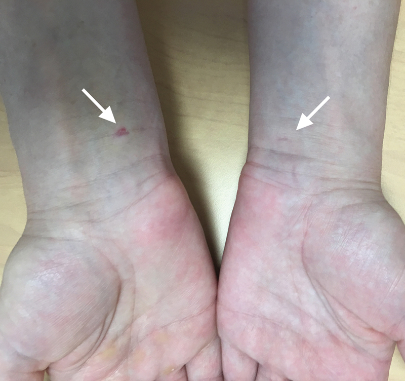
Carpal tunnel syndrome (CTS) may be treated surgically if medical treatment fails. The classical approach involves release of the flexor retinaculum by endoscopic or open surgery. Meta-analyses have shown that the risk of nerve injury may be higher with endoscopic treatment. The recent contribution of ultrasound to the diagnosis and therapeutic management of CTS opens new perspectives. Ultrasound-guided carpal tunnel release via a minimally invasive approach enables the whole operation to be performed as a percutaneous radiological procedure. The advantages are a smaller incision compared with classical techniques; great safety during the procedure by visualization of anatomic structures, particularly variations in the median nerve; and realization of the procedure under local anesthesia. These advantages lead to a reduction in postsurgical sequelae and more rapid resumption of daily activities and work. Dressings are removed by the third day postsurgery. Recent studies seem to confirm the medical, economic, and aesthetic benefits of this new approach.
Keywords: carpal tunnel release; carpal tunnel syndrome; interventional radiology; minimally invasive surgery; ultrasound-guided surgery; ultrasound-guided treatment.
Figures
Similar articles
-
Dynamic ultrasound for evaluating the adequacy of median nerve decompression following minimally invasive carpal tunnel release: technical innovation and case study.Heliyon. 2023 Jan 19;9(1):e13107. doi: 10.1016/j.heliyon.2023.e13107. eCollection 2023 Jan. Heliyon. 2023. PMID: 36711298 Free PMC article.
-
Treatment of carpal tunnel syndrome : from ultrasonography to ultrasound guided carpal tunnel release.Joint Bone Spine. 2018 Oct;85(5):545-552. doi: 10.1016/j.jbspin.2017.11.003. Epub 2017 Nov 16. Joint Bone Spine. 2018. PMID: 29154980 Review.
-
Carpal tunnel release using the KnifeLight technique: An alternative to endoscopic approach?Int J Surg Case Rep. 2024 Dec;125:110609. doi: 10.1016/j.ijscr.2024.110609. Epub 2024 Nov 15. Int J Surg Case Rep. 2024. PMID: 39550812 Free PMC article.
-
Minimally invasive carpal tunnel decompression using the KnifeLight.Neurosurgery. 2007 Feb;60(2 Suppl 1):ONS162-8; discussion ONS168-9. doi: 10.1227/01.NEU.0000249249.33052.7E. Neurosurgery. 2007. PMID: 17297379
-
Safe Zones for Percutaneous Carpal Tunnel Release.Hand Clin. 2022 Feb;38(1):83-90. doi: 10.1016/j.hcl.2021.08.008. Hand Clin. 2022. PMID: 34802612 Review.
Cited by
-
Percutaneous Ultrasound-Assisted Carpal Tunnel Release Using Sono-Instruments®.Cureus. 2024 Aug 14;16(8):e66899. doi: 10.7759/cureus.66899. eCollection 2024 Aug. Cureus. 2024. PMID: 39280410 Free PMC article.
-
Dynamic ultrasound for evaluating the adequacy of median nerve decompression following minimally invasive carpal tunnel release: technical innovation and case study.Heliyon. 2023 Jan 19;9(1):e13107. doi: 10.1016/j.heliyon.2023.e13107. eCollection 2023 Jan. Heliyon. 2023. PMID: 36711298 Free PMC article.
-
Prospective evaluation of a novel device for ultrasound-guided percutaneous treatment of carpal tunnel and trigger finger disease. Efficacy and safety of sono-instruments®.J Ultrasound. 2024 Dec;27(4):873-885. doi: 10.1007/s40477-023-00851-y. Epub 2024 Apr 10. J Ultrasound. 2024. PMID: 38600313 Free PMC article.
-
Variations of the Median Nerve and Carpal Tunnel Syndrome: a Systematic Review of the Literature.Maedica (Bucur). 2023 Dec;18(4):699-704. doi: 10.26574/maedica.2023.18.4.699. Maedica (Bucur). 2023. PMID: 38348062 Free PMC article.
-
Carpal tunnel ultrasound: is the "safe zone" on the ulnar side of the median nerve really avascular?Eur Radiol. 2020 Feb;30(2):887-894. doi: 10.1007/s00330-019-06416-0. Epub 2019 Aug 29. Eur Radiol. 2020. PMID: 31468160
References
-
- Marshall S, Tardif G, Ashworth N. Local corticosteroid injection for carpal tunnel syndrome. Cochrane Database Syst Rev. 2007;(02):CD001554. - PubMed
-
- Nakamichi K, Tachibana S. Ultrasonographically assisted carpal tunnel release. J Hand Surg Am. 1997;22(05):853–862. - PubMed
-
- Lecoq B, Hanouz N, Morello R et al.Ultrasound-assisted surgical release of carpal tunnel syndrome: results of a pilot open-label uncontrolled trial conducted outside the operating theatre. Joint Bone Spine. 2015;82(06):442–445. - PubMed
-
- Chern T C, Kuo L C, Shao C J, Wu T T, Wu K C, Jou I M. Ultrasonographically guided percutaneous carpal tunnel release: early clinical experiences and outcomes. Arthroscopy. 2015;31(12):2400–2410. - PubMed
-
- Capa-Grasa A, Rojo-Manaute J M, Rodríguez F C, Martín J V. Ultra minimally invasive sonographically guided carpal tunnel release: an external pilot study. Orthop Traumatol Surg Res. 2014;100(03):287–292. - PubMed
Publication types
LinkOut - more resources
Full Text Sources
Research Materials
