Review
. 2018 Oct;35(4):309-323.
doi: 10.1055/s-0038-1673639. Epub 2018 Nov 5.

Percutaneous Vertebral Augmentation Techniques in Osteoporotic and Traumatic Fractures

Affiliations
Review

Percutaneous Vertebral Augmentation Techniques in Osteoporotic and Traumatic Fractures

Valérie Bousson et al. Semin Intervent Radiol. 2018 Oct.

Abstract

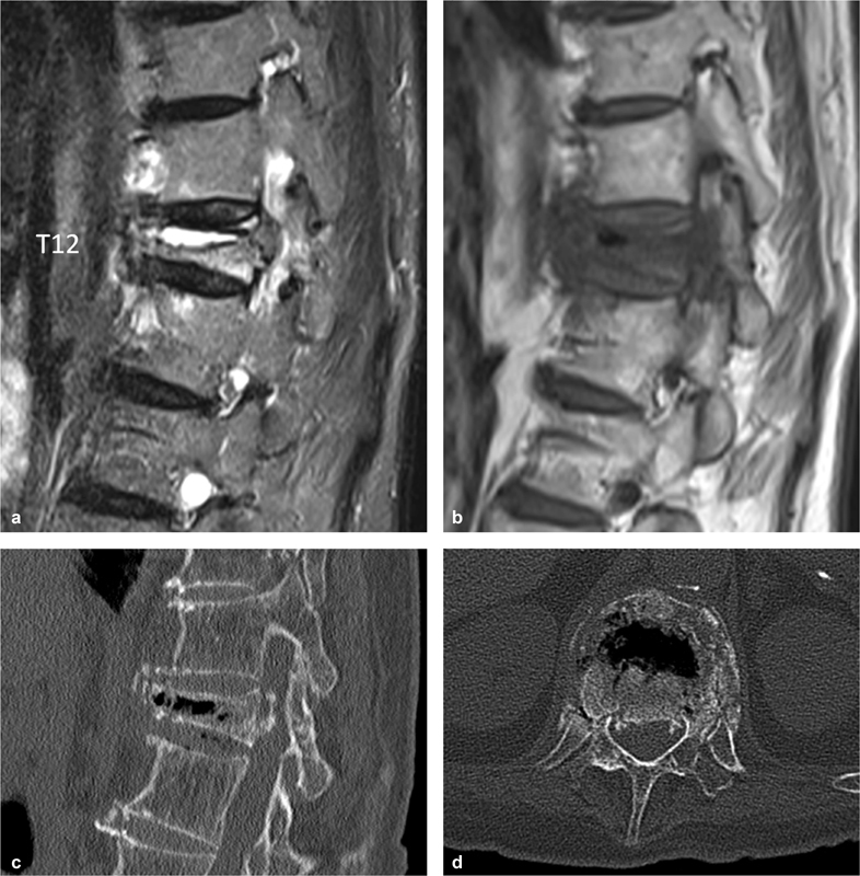
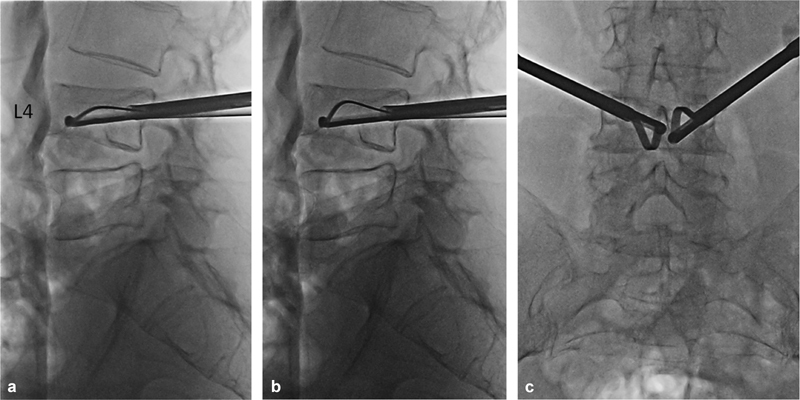
Percutaneous vertebral augmentation/consolidation techniques are varied. These are vertebroplasty, kyphoplasty, and several methods with percutaneous introduction of an implant (associated or not with cement injection). They are proposed in painful osteoporotic vertebral fractures and traumatic fractures. The objectives are to consolidate the fracture and, if possible, to restore the height of the vertebral body to reduce vertebral and regional kyphosis. Stabilization of the fracture leads to a reduction in pain and thus restores the spinal support function as quickly as possible, which is particularly important in the elderly. The effectiveness of these interventions on fracture pain was challenged once by two randomized trials comparing vertebroplasty to a sham intervention. Since then, many other randomized studies in support of vertebroplasty efficacy have been published. International recommendations reserve vertebroplasty for medical treatment failures on pain, but earlier positioning may be debatable if the objective is to limit kyphotic deformity or even reexpand the vertebral body. Recent data suggest that in osteoporotic fracture, the degree of kyphosis reduction achieved by kyphoplasty and percutaneous implant techniques, compared with vertebroplasty, is not sufficient to justify the additional cost and the use of a somewhat longer and traumatic procedure. In young patients with acute traumatic fractures and a significant kyphotic angle, kyphoplasty and percutaneous implant techniques are preferred to vertebroplasty, as in these cases a deformity reduction has a significant positive impact on the clinical outcome.

Keywords: interventional radiology; kyphoplasty; vertebral augmentation techniques; vertebral fracture; vertebroplasty.


Figures

Fig. 1
Fig. 1
Time course of an osteoporotic L1 fracture in a 72-year-old patient. On the initial T1-weighted MRI ( a ), the regional kyphosis angle was 17 degrees. The treatment was medical. The patient had an MRI 6 months later (b) for persistent thoracolumbar junction pain. The fracture increased, especially anteriorly, and the regional kyphosis angle increased from 17 to 22 degrees in the supine position (b).
Fig. 2
Fig. 2
Osteoporotic fracture of T12 in a 78-year-old patient. Intracorporeal fluid cavity on T2-weighted MRI ( a ), mixed liquid and gas on T1-weighted MRI (b), and gas on CT scan ( c and d ). This cavity corresponds to a pseudarthrosis.
Fig. 3
Fig. 3
Cascade of osteoporotic fractures in a 76-year-old patient. The initial MRI ( a and b ) shows a recent or semi-recent fracture of the T10 and L2 vertebrae, and a discrete fracture of the lower L4 endplate. The persistence of pain leads the physician to propose a vertebroplasty. The new MRI ( c and d ), performed 2 months after the first one, shows four new fractures, T11, T12, upper endplate of L4, and L5.
Fig. 4
Fig. 4
T12 fracture with marked regional kyphosis. CT, sagittal image ( a ); radiography in flexion (b) .
Fig. 5
Fig. 5
L1 vertebral kyphoplasty in a 68-year-old patient. The fractured L1 vertebra is the site of a pseudarthrosis on the T2 sagittal sequence ( a ) and radiograph ( b ). The steps of the intervention are introduction of the trocars ( c ), introduction of the drill ( d ), introduction of the balloons ( e ), the balloons are inflated ( f , g ) and then removed. Cement injection gradually filled the cavity ( h ). Control X-rays ( i and j ), and CT ( k and l ).
Fig. 6
Fig. 6
( a–c ) L4 vertebral fracture and reduction instruments. Gradual spreading of the slats under the upper left and lower right endplates.
Fig. 7
Fig. 7
Kyphoplasty and percutaneous fixation for a traumatic fracture of L4 in a 55-year-old patient. The radiograph ( a ) and the CT scan ( b ) show a fracture of L4, type Magerl A3, with regional traumatic angulation of about 30 degrees. Partial restoration of vertebral body height by balloon kyphoplasty ( c ). Radiographs ( d, e ) showing the result of kyphoplasty coupled with percutaneous fixation L3–L5, with restoration of a relatively satisfactory lumbar lordosis.
Fig. 8
Fig. 8
L1 vertebral body height gain after cementoplasty of a traumatic fracture less than 1 week old in a 74-year-old patient. ( a–c ) X-ray and CT scan immediately before cementoplasty; ( d–f ) X-ray and CT scan immediately after cementoplasty.
Fig. 9
Fig. 9
Vertebroplasty of L1 and L2 vertebrae site of osteoporotic fractures in a 78-year-old male with Parkinson's disease. MRI ( a ) shows a large pseudarthrosis cavity. The X-ray before cementoplasty shows the reduced vertebral body height of L1 ( b ). After insertion of the trocars ( c ), when the L1 mandrel was removed, the cavity was filled with gas ( d ). L1 and L2 cementoplasty with L1 vertebral body height gain ( e and f ).
Fig. 10
Fig. 10
Successive fractures and vertebroplasties in an 87-year-old male. Osteoporotic fracture of L1 ( a ) treated with cementoplasty ( b, c ); fracture of T12 (vertebra above cementoplasty) 1 month after L1 cementoplasty ( d ); cementoplasty of T12 ( e ); fracture of T11 (vertebra above cementoplasty) 3 weeks after T12 cementoplasty ( f ).
Fig. 11
Fig. 11
Successive fractures and vertebroplasties in a 68-year-old female. Fractures of T11 and L1 ( a ); vertebroplasty of T11, L1, and T12 preventively ( b ); 15 days later, occurrence of a fracture of T10 (adjacent to vertebroplasty) and L3 (distant from vertebroplasty) ( c ).

Similar articles

Cited by

References

    1. Fechtenbaum J, Cropet C, Kolta S, Verdoncq B, Orcel P, Roux C. Reporting of vertebral fractures on spine X-rays. Osteoporos Int. 2005;16(12):1823–1826. - PubMed
    1. Lindsay R, Silverman S L, Cooper C et al.Risk of new vertebral fracture in the year following a fracture. JAMA. 2001;285(03):320–323. - PubMed
    1. Roux C, Fechtenbaum J, Kolta S, Said-Nahal R, Briot K, Benhamou C-L. Prospective assessment of thoracic kyphosis in postmenopausal women with osteoporosis. J Bone Miner Res. 2010;25(02):362–368. - PubMed
    1. Edidin A A, Ong K L, Lau E, Kurtz S M. Life expectancy following diagnosis of a vertebral compression fracture. Osteoporos Int. 2013;24(02):451–458. - PubMed
    1. Magerl F, Aebi M, Gertzbein S D, Harms J, Nazarian S. A comprehensive classification of thoracic and lumbar injuries. Eur Spine J. 1994;3(04):184–201. - PubMed