. 2018 Oct 26;18(5):e37.
doi: 10.4110/in.2018.18.e37. eCollection 2018 Oct.

Oral Administration of Silk Peptide Enhances the Maturation and Cytolytic Activity of Natural Killer Cells

Affiliations

Oral Administration of Silk Peptide Enhances the Maturation and Cytolytic Activity of Natural Killer Cells

Sun-Hee Jang et al. Immune Netw. .

Abstract

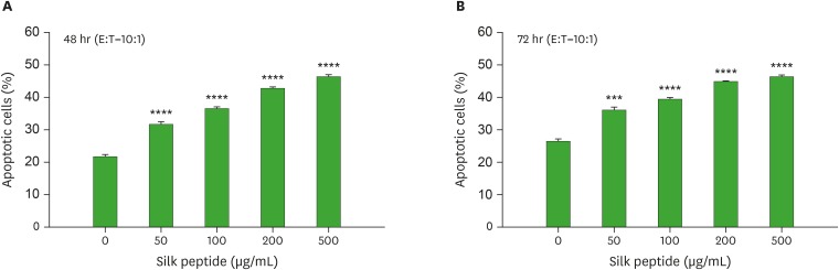
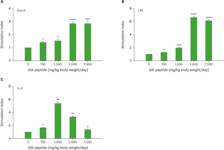
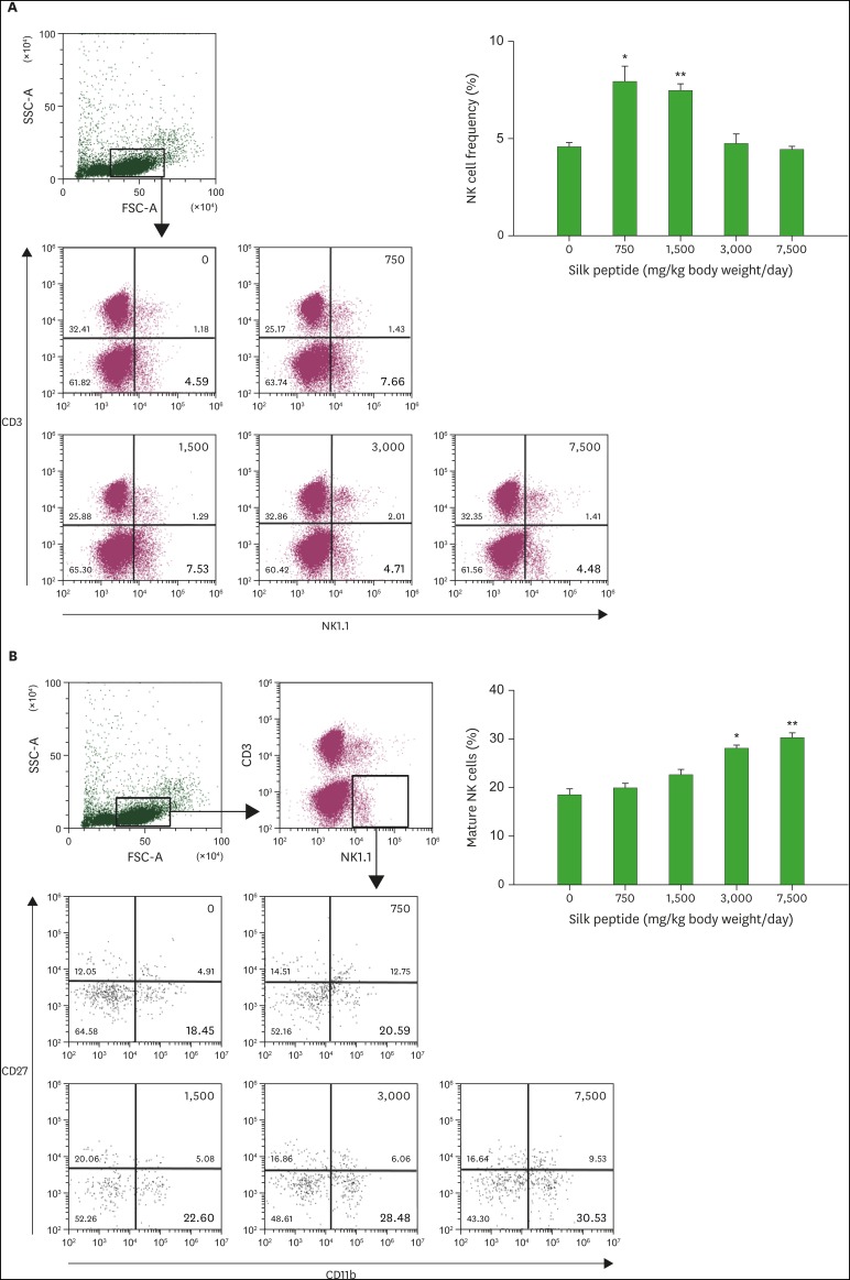
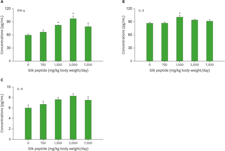
Silk peptide, the hydrolysate of silk protein derived from cocoons, has been employed as a biomedical material and is believed to be safe for human use. Silk peptide display various bioactivities, including anti-inflammatory, immune-regulatory, anti-tumor, anti-viral, and anti-bacterial. Although earlier investigations demonstrated that silk peptide stimulates macrophages and the production of pro-inflammatory cytokines, its effect on natural killer (NK) cell function has not yet been explored. In this study, we initially confirmed that silk peptide enhances NK cell activity in vitro and ex vivo. To assess the modulatory activity of silk peptide on NK cells, mice were fed various amounts of a silk peptide-supplemented diet for 2 months and the effects on immune stimulation, including NK cell activation, were evaluated. Oral administration of silk peptide significantly enhanced the proliferation of mitogen- or IL-2-stimulated splenocytes. In addition, oral silk peptide treatment enhanced the frequency and degree of maturation of NK cells in splenocytes. The same treatment also significantly enhanced the target cell cytolytic activity of NK cells, which was determined by cell surface CD107a expression and intracellular interferon-γ expression. Finally, oral administration of silk peptide stimulated T helper 1-type cytokine expression from splenic lymphocytes. Collectively, our results suggest that silk peptide potentiates NK cell activity in vivo and could be used as a compound for immune-modulating anti-tumor treatment.

Keywords: Cytokine; Cytolytic activity; In vivo; Natural killer cells; Silk peptide.


Conflict of interest statement

Conflict of Interest: The authors declare no potential conflict of interest.

Figures

Figure 1
Figure 1. Effect of ex vivo silk peptide treatment of splenocytes on NK cell cytolytic activity against YAC-1 target cells. Splenocytes were incubated with the indicated concentrations of silk peptide for (A) 48 h or (B) 72 h and incubated with PKH-26-labeled YAC-1 target cells at a ratio of 10:1. The degree of target cell lysis was measured as described in the Materials and Methods. Data are presented as the mean±SE (n=3) of three independent experiments.
E:T, effector to target ratio. ***p<0.001, ****p<0.0001 indicate a significant difference compared to the control.
Figure 2
Figure 2. Influence of oral administration of silk peptide on mitogen- or IL-2-mediated proliferation of splenocytes. Mice were orally administered various amounts of silk peptide-supplemented feed for 2 months and splenocytes were prepared. The level of (A) Con A-, (B) LPS-, or (C) IL-2-mediated proliferation of splenocytes was measured as described in the Materials and Methods. Data are presented as the mean±SE (n=3) of three independent experiments.
*p<0.05, **p<0.01, ***p<0.001, and ****p<0.0001 indicate a significant difference compared to the control (without silk peptide treatment).
Figure 3
Figure 3. Influence of oral administration of silk peptide on NK cell frequency and the status of NK cell maturation in splenocytes. Changes in (A) NK cell frequency (CD3NK1.1+) and (B) mature NK cells (CD11bhighCD27low) in splenocytes prepared from mice orally administered various amount of silk peptide were analyzed as described in the Materials and Methods. FACS plot shows a representative result and data shown in bar graph are presented as the mean±SE (n=3) of three independent experiments.
FSC-A, forward-scatter area; SSC-A, side-scatter area. *p<0.05 and **p<0.01 indicate a significant difference compared to the control (without silk peptide treatment).
Figure 4
Figure 4. Cytolytic activity of NK cells purified from splenocytes. NK cells were purified from splenocytes prepared from mice fed various amounts of silk peptide and stimulated with YAC-1 target cells at a ratio of 5:1 in the presence of brefeldin A and monensin. NK cell cytolytic activity was determined by measuring (A) cell surface CD107a expression and (B) intracellular IFN-γ expression as described in the Materials and Methods. FACS plot shows a representative result and data shown in bar graph are presented as the mean±SE (n=3) of three independent experiments. NC and PC show the minimum and maximum levels, respectively.
FSC-A, forward-scatter area; SSC-A, side-scatter area; NC, negative control (unstimulated control containing NK cell only without target cells); PC, positive control (NK cells stimulated with PMA and ionomycin). *p<0.05 and **p<0.01 indicate a significant difference compared to the control (without silk peptide treatment).
Figure 5
Figure 5. Level of cytokine expression following stimulation of splenocytes prepared from mice fed various amount of silk peptide with PMA and ionomycin. Splenocytes prepared from silk peptide-treated mice were stimulated in vitro with PMA and ionomycin. The expression levels of the Th1-type cytokines (A) IFN-γ and (B) IL-2, and the Th2-type cytokine (C) IL-4 were determined as described in the Materials and Methods. Data are presented as the mean±SE (n=3) of three independent experiments.
*p<0.05 indicates a significant difference compared to the control (without silk peptide treatment).

Similar articles

Cited by

References

    1. Mandal A, Viswanathan C. Natural killer cells: in health and disease. Hematol Oncol Stem Cell Ther. 2015;8:47–55. - PubMed
    1. Moretta L, Ciccone E, Moretta A, Höglund P, Ohlén C, Kärre K. Allorecognition by NK cells: nonself or no self? Immunol Today. 1992;13:300–306. - PubMed
    1. Vivier E, Tomasello E, Baratin M, Walzer T, Ugolini S. Functions of natural killer cells. Nat Immunol. 2008;9:503–510. - PubMed
    1. Glas R, Franksson L, Une C, Eloranta ML, Ohlén C, Örn A, Kärre K. Recruitment and activation of natural killer (NK) cells in vivo determined by the target cell phenotype. An adaptive component of NK cell-mediated responses. J Exp Med. 2000;191:129–138. - PMC - PubMed
    1. Smyth MJ, Crowe NY, Hayakawa Y, Takeda K, Yagita H, Godfrey DI. NKT cells - conductors of tumor immunity? Curr Opin Immunol. 2002;14:165–171. - PubMed