. 2018 Oct 9:2018:5106174.
doi: 10.1155/2018/5106174. eCollection 2018.

Excess Hydrocortisone Hampers Placental Nutrient Uptake Disrupting Cellular Metabolism

Affiliations

Excess Hydrocortisone Hampers Placental Nutrient Uptake Disrupting Cellular Metabolism

Rosa María Mateos et al. Biomed Res Int. .

Abstract

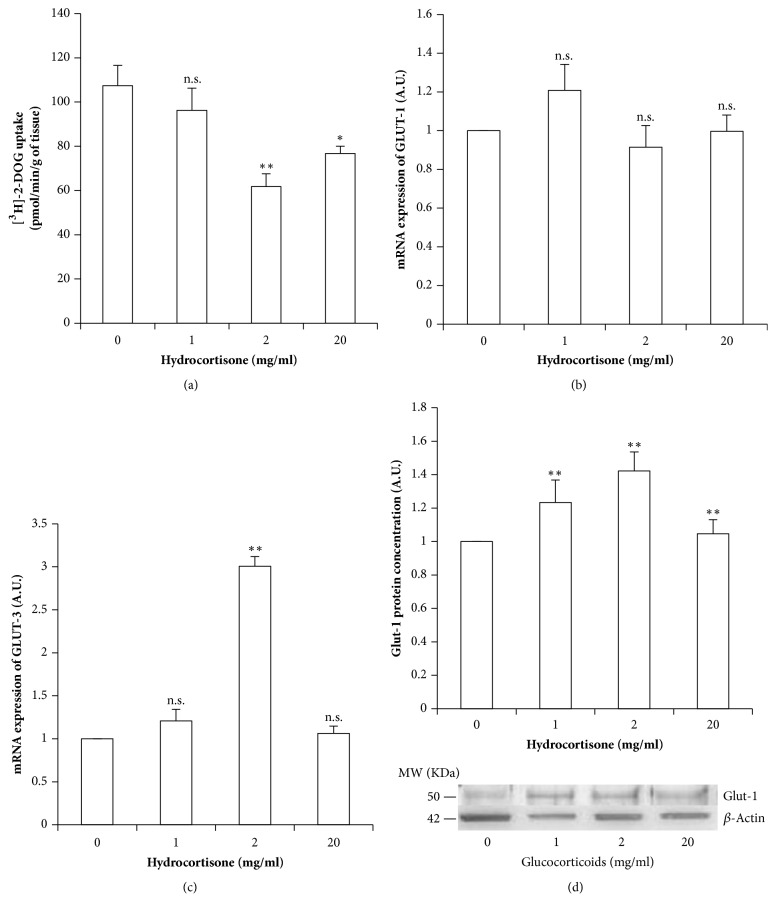
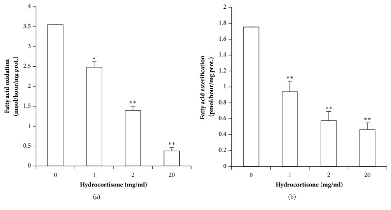
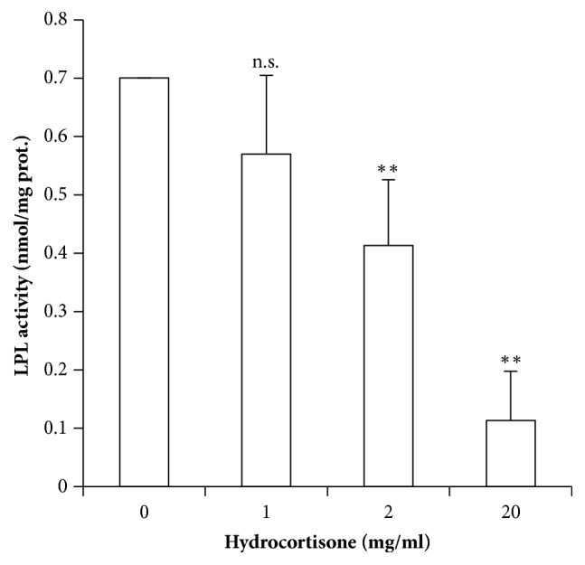
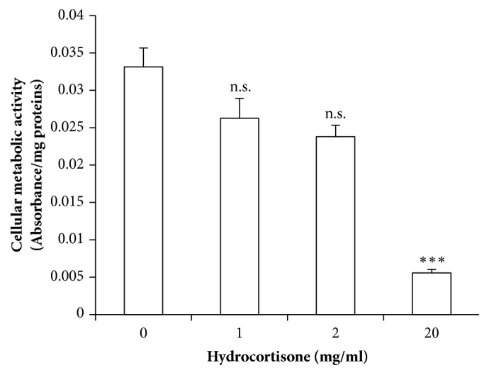
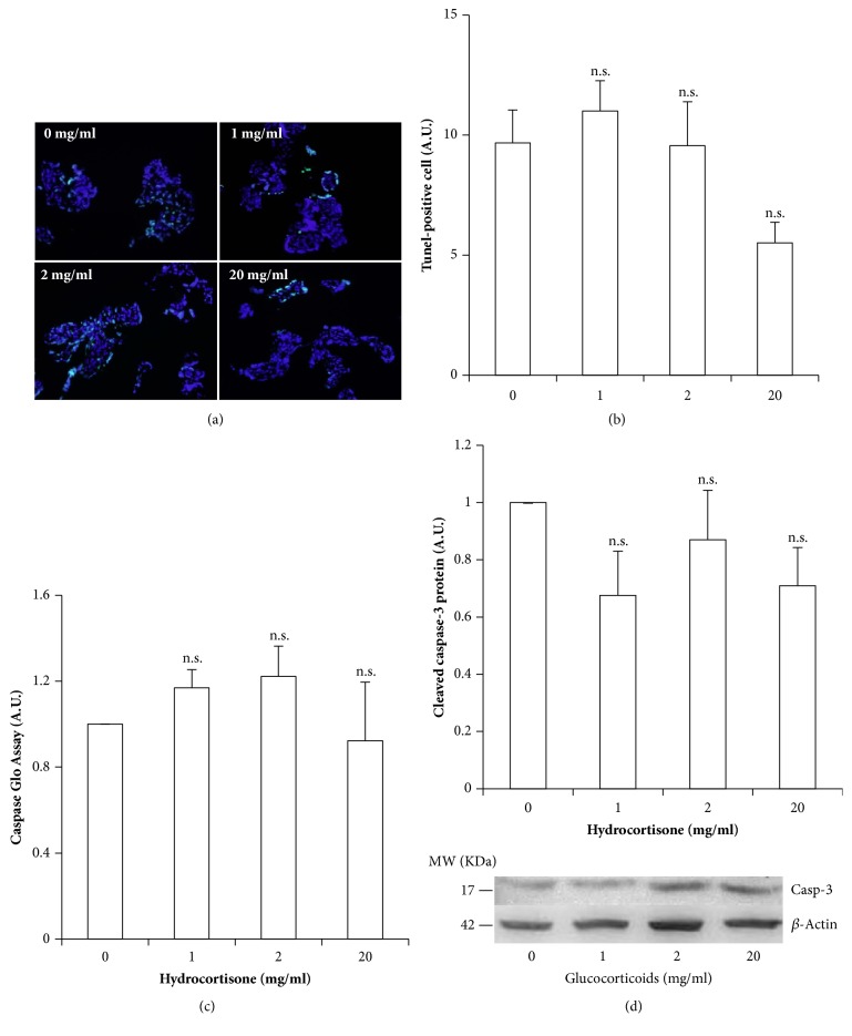
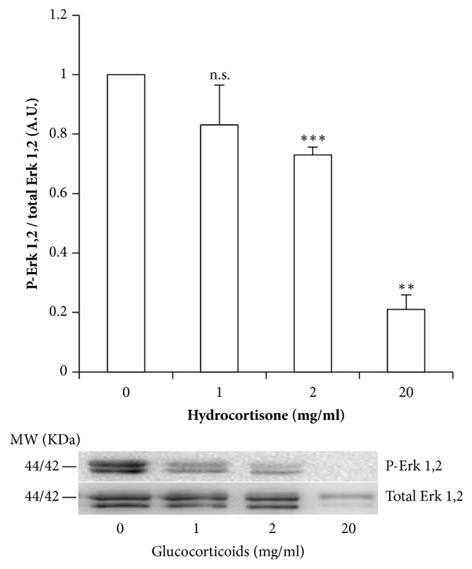
Low birth weight increases neonatal morbidity and mortality, and surviving infants have increased risk of metabolic and cardiovascular disturbances later in life, as well as other neurological, psychiatric, and immune complications. A gestational excess of glucocorticoids (GCs) is a well-known cause for fetal growth retardation, but the biological basis for this association remains elusive. Placental growth is closely related to fetal growth. The placenta is the main regulator of nutrient transport to the fetus, resulting from the difference between placental nutrient uptake and the placenta's own metabolism. The aim of this study was to analyze how excess hydrocortisone affects placental glucose and lipid metabolism. Human placenta explants from term physiological pregnancies were cultured for 18 hours under different hydrocortisone concentrations (2.75, 5.5, and 55 mM; 1, 2, and 20 mg/ml). Placental glucose and lipid uptake and the metabolic partitioning of fatty acids were quantified by isotopic techniques, and expression of specific glucose transporter GLUT1 was quantified by western blot. Cell viability was assessed by MTT, immunohistochemistry and caspase activity. We found that excess hydrocortisone impairs glucose uptake and lipoprotein lipase (LPL) activity, coincident with a GC-dose dependent inhibition of fatty acid oxidation and esterification. None of the experimental conditions showed an increased cell death. In conclusion, our results show that GC overexposure exerts a dysfunctional effect on lipid transport and metabolism and glucose uptake in human placental explants. These findings could well be directly related to a reduced placental growth and possibly to a reduced supply of nutrients to the fetus and the consequent fetal growth retardation and metabolic programming.


Figures

Figure 1
Figure 1
Hydrocortisone's effects on glucose uptake and glucose transporters in placental explants. (a) Glucose uptake as measured by [3H]-2-DOG incorporation. (b) GLUT1 mRNA expression normalized by actin mRNA expression. (c) GLUT3 mRNA expression normalized by actin mRNA. (d) GLUT1 protein levels as measured by western blot, normalized by actin levels, together with a representative photograph. Values are represented as means ± SEM for 5 independent experiments. p<0.05; ∗∗p<0.025; ∗∗∗p<0.01 relative to control (A.U.: arbitrary units).
Figure 2
Figure 2
Hydrocortisone's effects in placental explants' lipid metabolism. (a) Fatty acid oxidation, in nmol/hour/mg of protein, was inhibited by glucocorticoids in a dose dependent manner. (b) Fatty acid esterification, in pmol/hour/mg of protein, was also decreased in a dose dependent manner. Values are represented as means ± SEM for at least 5 independent experiments, in triplicate. p≤0.05; ∗∗p≤0.01; ∗∗∗p≤0.001 relative to control.
Figure 3
Figure 3
LPL activity of placental explants with increasing hydrocortisone concentrations. Values measured in nmol/mg of proteins are represented as means ± SEM for at least 4 independent experiments in triplicate. p≤0.05; ∗∗p≤0.01; ∗∗∗p≤0.001 relative to control.
Figure 4
Figure 4
Mitochondrial activity as measured by MTT. Absorbance per mg of protein content, represented as means ± SEM for 6 independent experiments. p<0.05; ∗∗p<0.025; ∗∗∗p<0.01 relative to control.
Figure 5
Figure 5
Assessment of hydrocortisone-induced cellular death by apoptosis in healthy-term placental explants. (a) Representative microphotographs of TUNEL assay to determine apoptosis in each experimental condition; cells undergoing apoptosis present fluorescent green nuclei. (b) Quantification of the % of TUNEL positive cells per condition. (c) Active caspases 3 and 7 as measured by luminometry, using the Caspase Glo Assay. (d) Cleaved caspase 3 as determined by western blot and normalized by actin levels. Values are presented as means ± SEM for 5 independent experiments. p≤0.05; ∗∗p≤0.01; ∗∗∗p≤0.001 relative to control.
Figure 6
Figure 6
Dose dependent inhibition of MAPK-signaling pathway in placental explants by hydrocortisone. Western blot analysis of P-Erk1,2 vs. Total Erk1,2. Values are means ± SEM for 6 independent experiments. p<0.05; ∗∗p<0.025; ∗∗∗p<0.010 relative to control.

Similar articles

Cited by

References

    1. Barker D. J. P. The developmental origins of adult disease. Journal of the American College of Nutrition. 2004;23:588–595. doi: 10.1080/07315724.2004.10719428. - DOI - PubMed
    1. Vitale G., Salvioli S., Franceschi C. Oxidative stress and the ageing endocrine system. Nature Reviews Endocrinology. 2013;9(4):228–240. doi: 10.1038/nrendo.2013.29. - DOI - PubMed
    1. Battaglia F. C., Meschia G. Fetal Nutrition. Annual Review of Nutrition. 1988;8(1):43–61. doi: 10.1146/annurev.nu.08.070188.000355. - DOI - PubMed
    1. Jones H. N., Powell T. L., Jansson T. Regulation of placental nutrient transport - a review. Placenta. 2007;28(8-9):763–774. doi: 10.1016/j.placenta.2007.05.002. - DOI - PubMed
    1. Brett K. E., Ferraro Z. M., Yockell-Lelievre J., Gruslin A., Adamo K. B. Maternal–fetal nutrient transport in pregnancy pathologies: the role of the placenta. International Journal of Molecular Sciences. 2014;15(9):16153–16185. doi: 10.3390/ijms150916153. - DOI - PMC - PubMed

MeSH terms