

Automatic Global Level Set Approach for Lumbar Vertebrae CT Image Segmentation
- PMID: 30402488
- PMCID: PMC6196995
- DOI: 10.1155/2018/6319879
Automatic Global Level Set Approach for Lumbar Vertebrae CT Image Segmentation
Abstract
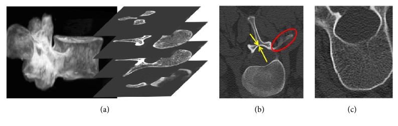
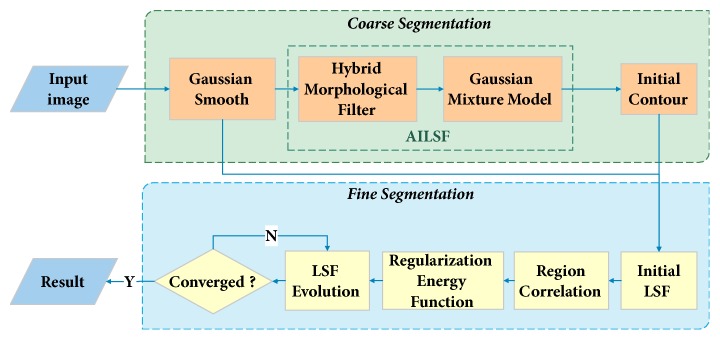
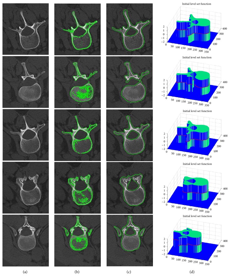
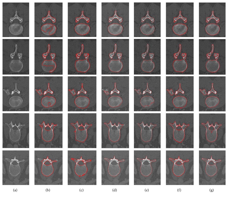
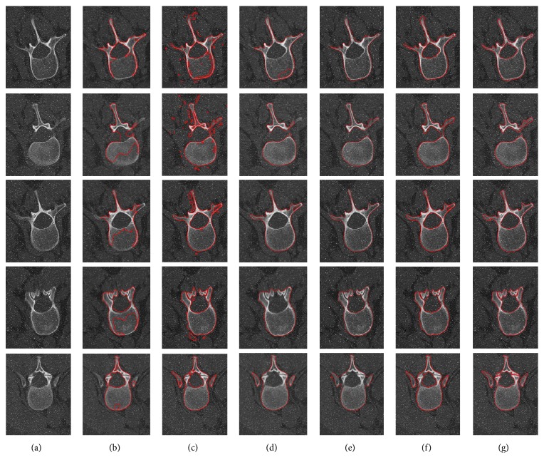
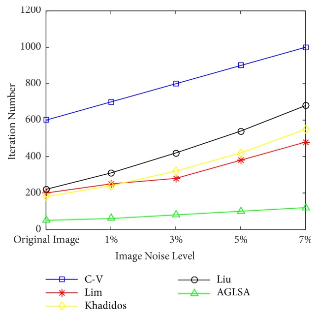
Vertebrae computed tomography (CT) image automatic segmentation is an essential step for Image-guided minimally invasive spine surgery. However, most of state-of-the-art methods still require human intervention due to the inherent limitations of vertebrae CT image, such as topological variation, irregular boundaries (double boundary, weak boundary), and image noise. Therefore, this paper intentionally designed an automatic global level set approach (AGLSA), which is capable of dealing with these issues for lumbar vertebrae CT image segmentation. Unlike the traditional level set methods, we firstly propose an automatically initialized level set function (AILSF) that comprises hybrid morphological filter (HMF) and Gaussian mixture model (GMM) to automatically generate a smooth initial contour which is precisely adjacent to the object boundary. Secondly, a regularized level set formulation is introduced to overcome the weak boundary leaking problem, which utilizes the region correlation of histograms inside and outside the level set contour as a global term. Ultimately, a gradient vector flow (GVF) based edge-stopping function is employed to guarantee a fast convergence rate of the level set evolution and to avoid level set function oversegmentation at the same time. Our proposed approach has been tested on 115 vertebrae CT volumes of various patients. Quantitative comparisons validate that our proposed AGLSA is more accurate in segmenting lumbar vertebrae CT images with irregular boundaries and more robust to various levels of salt-and-pepper noise.
Figures

References
-
- Kadoury S., Labelle H., Parent S. Medical Image Computing and Computer-Assisted Intervention – MICCAI 2014. Vol. 8675. Cham: Springer International Publishing; 2014. 3D Spine Reconstruction of Postoperative Patients from Multi-level Manifold Ensembles; pp. 361–368. (Lecture Notes in Computer Science). - DOI - PubMed
-
- Nitin A., Daniel P. F., Raghav G., et al. A comparative analysis of minimally invasive and open spine surgery patient education resources. Journal of Neurosurgery: Spine. 2014;21(3):468–474. - PubMed
-
- Aslan M. S., Shalaby A., Farag A. A. Vertebral body segmentation using a probabilistic and universal shape model. IET Computer Vision. 2015;9(2):234–250. doi: 10.1049/iet-cvi.2013.0154. - DOI
MeSH terms
LinkOut - more resources
Full Text Sources
Medical
