. 2018 Oct 31;4(10):eaat5118.
doi: 10.1126/sciadv.aat5118. eCollection 2018 Oct.

Organic synthesis on Mars by electrochemical reduction of CO2

Affiliations

Organic synthesis on Mars by electrochemical reduction of CO2

A Steele et al. Sci Adv. .

Abstract

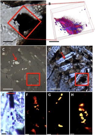
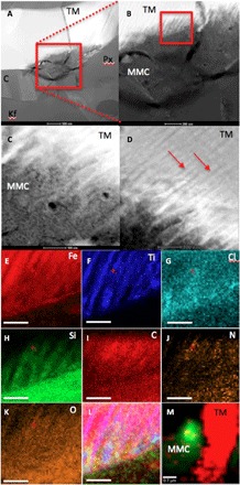
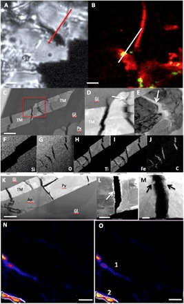
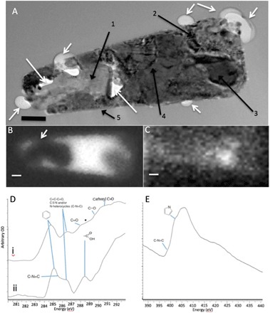
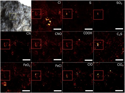
The sources and nature of organic carbon on Mars have been a subject of intense research. Steele et al. (2012) showed that 10 martian meteorites contain macromolecular carbon phases contained within pyroxene- and olivine-hosted melt inclusions. Here, we show that martian meteorites Tissint, Nakhla, and NWA 1950 have an inventory of organic carbon species associated with fluid-mineral reactions that are remarkably consistent with those detected by the Mars Science Laboratory (MSL) mission. We advance the hypothesis that interactions among spinel-group minerals, sulfides, and a brine enable the electrochemical reduction of aqueous CO2 to organic molecules. Although documented here in martian samples, a similar process likely occurs wherever igneous rocks containing spinel-group minerals and/or sulfides encounter brines.


Figures

Fig. 1
Fig. 1. Light and CRIS of the relationship between magnetite and MMC in the meteorites studied.
(A) Transmitted light microscopy image of a darkened area within maskelynite, entrained in the subsurface to the thin section in NWA 1950 (scale bar, 100 μm). The red box indicates the area for three-dimensional (3D) mapping by CRIS. (B) A 3D depth profile composite CRIS image of magnetite (red) and MMC (blue) (slices are 2 μm apart) from the area denoted by the red box in (A) (scale bar, ~60 μm). (C and D) The same area of Nakhla mesostasis imaged in transmission and reflection showing a magnetite grain beneath the surface of the section (marked by red arrows) (scale bars, 20 μm). (E) Transmitted light image of a magnetite-rich area (dark vertical band) in the Tissint meteorite [scale bar, 20 μm; red line on top delineates the area where a focused ion beam (FIB) section was removed for analyses; see Fig. 4]. (F to H) CRIS imaging maps taken at 8 μm into the surface of the thin section and depth profile of the feature shown in (A); (F) magnetite, (G) pyrite, and (H) MMC.
Fig. 2
Fig. 2. High-resolution TEM analysis of magnetite and MMC features within the Nakhla meteorite.
(A to D) High-angle annular dark-field (HAADF) TEM images showing (A) a Nakhla titano-magnetite (TM) grain extracted through FIB milling from the area marked with a red box in Fig. 1 (C and D). The grain is surrounded by pyroxene (Px), K-feldspar (Kf), and cristobalite (C) (scale bar, 500 nm). (B) A higher-magnification image of the red boxed area in (A), showing evidence of corrosion with a comb-like etching pattern into the TM grain (TM; scale bar, 200 nm). (C) A higher-magnification and high-contrast image of the area denoted by the red box in (B), with the titanium-rich exsolution lamellae in the TM corroded grain visible (red arrows; scale bar, 100 nm). (D) Area showing the interaction with TM exsolution lamellae and the surrounding matrix; the titanium-rich lamellae are highlighted with red arrows; EDX and selected-area electron diffraction (SAED) analyses of this matrix revealed a carbon-rich but amorphous (no diffraction contrast detected during tilting of the sample) assemblage (MMC; scale bar, 100 nm). (E to K) X-ray intensity maps of the exsolution features from the red box in (B), showing the distribution and associations between (scale bars, 100 nm). (E) Iron, (F) titanium, (G) chlorine, (H) silicon, (I) carbon, (J) nitrogen, and (K) oxygen, while (L) is a three-color composite map of Fe (red), Ti (blue), and Cl (green) to show the link between the corrosion of the TM grain and a chlorine-rich brine. (M) Composite CRIS map of the FIB section after TEM analysis showing titano-magnetite (red) and MMC (green), confirming the presence of MMC before and after analysis by TEM [note that map (M) is rotated and at a lower magnification compared with the x-ray maps in (A) to (L) and fig. S1; scale bar, 0.7 μm].
Fig. 3
Fig. 3. TEM and hydrogen isotopic analysis of MMC-rich areas in the Tissint meteorite.
(A) Light microscopy image of a TM lath within maskelynite in the Tissint meteorite (dark area across the line denotes the area from which the FIB section was extracted). Scale bar, 3 μm. (B) Composite CRIS map showing TM in red and MMC in green; the white line corresponds to the red FIB line in (A). Scale bar, 3 μm. (C to E) TEM HAADF images of the extracted FIB section shown in (A) and (B), illustrating that the TM lath is cracked along its length. Scale bars, 500 nm (C) and 100 nm (D and E). (D) A closeup image of the red box in (C), showing two of the cracks and evidencing that the edge of the cracks are corroded (white arrow). (E) Cracks filled with MMC as documented in the EDX maps in (F) to (J). (F) Silicon, (G) oxygen, (H) titanium, (I) iron, and (J) carbon. Scale bars, 1 μm (F to J). (K to M) HAADF image of an inclusion assemblage in NWA 1950. The high-resolution images and analyses of the FIB section revealed that TM, and in this case, pyrrhotite and apatite laths are also cracked, and we know from the CRIS mapping shown in Fig. 1B that the magnetite is intimately interlinked in NWA 1950 with MMC. Scale bars, 2 μm (K), 300 nm (L), and 200 nm (M). Crack in pyrrhotite with the edges lined with deposited magnetite nanocrystals (area of high brightness lining the crack marked with a white arrows). (M) Close-up of the TM grains again showing evidence of corrosion along the edges of the cracks in the grains (arrows). (N) C13, nanoSIMS map of a different TM lath in the Tissint meteorite that had previously been confirmed by CRIS to contain MMC. Scale bar, 5 μm. (O) Deuterium map of the same area as in (N); area 1 recorded a δD of 2252 ± 392‰ and area 2 recorded a δD of 4536 ± 1113‰, scale bar, 5 μm.
Fig. 4
Fig. 4. TEM imaging and soft transmission x-ray analysis of MMC within an inclusion of the Tissint meteorite.
(A) Bright-field TEM image of a FIB section from the Tissint meteorite milled from the area analyzed by CRIS. The image reveals a highly complex inclusion. White arrows indicate bubbles protruding into the surrounding maskelynite matrix or void space inside the inclusion. Black arrows point to specific mineral phases identified by high-resolution imaging and EDX and SAED analyses (figs. S2 and S3) corresponding to (1) anhydrite, (2) magnetite and carbon, (3) Ni-containing pyrrhotite, (4) nanocrystalline multiphase magnetite and calcium-aluminium silicate phases, and (5) rim of sheet silicate–like aluminosilicate, probably montmorillonite (scale bar, 150 nm). (B) STXM map of Fe at 708.62 eV showing the iron-rich area of the inclusion (in white), including the area around the anhydrite grain and the bubble projecting into the glass matrix (arrow). (C) STXM map at the C-K edge at 284.9 eV showing a hot spot of aromatic carbon corresponding to pyrrhotite and magnetite (areas 2 and 3) shown in (A) (lighter shade shows greater concentration). Scale bars, 200 nm. (D) STXM spectrum of two inclusions from Tissint: (i) hot spot area in (C), showing the presence of peaks at 284.9, 286.4, and 288.56 eV, with a star demarking a possible shoulder peak at 288.56 eV; (ii) peaks from another inclusion containing many of the same functional groups as in (i) but also a shoulder at 285.6 eV, which is possibly indicative of 400 eV and pyrrole functionality at 405 eV. (E) STXM N-K edge spectrum from the same inclusion as spectrum (i) in (D), confirming the presence of C–N=C functionality at 400 eV and a pyrrole group at 405 eV. OD, optical density.
Fig. 5
Fig. 5. ToF-SIMS analysis of fresh fracture surface of the Tissint meteorite.
The first image is a light microscope image of a fresh fracture surface of the Tissint meteorite, as analyzed within the ToF-SIMS instrument. Individual species are recorded in the top right of the individual images and include Cl, S, SO3, CN, CNO, COOH, C2S, FeO2, FeCl, ClO, and ClO4. The red box in the first ion image of the first row (Cl) denotes an area of concentration of many carbon-, nitrogen-, chlorine-, iron-, and sulfur-containing species that are labeled in the top right of the remaining images. Scale bars, 20 μm. See table S2 for absolute peak assignments and fig. S5 for further species maps.

References

    1. Steele A., McCubbin F. M., Fries M., Kater L., Boctor N. Z., Fogel M. L., Conrad P. G., Glamoclija M., Spencer M., Morrow A. L., Hammond M. R., Zare R. N., Vicenzi E. P., Siljeström S., Bowden R., Herd C. D. K., Mysen B. O., Shirey S. B., Amundsen H. E. F., Treiman A. H., Bullock E. S., Jull A. J. T., A reduced organic carbon component in martian basalts. Science. 337, 212–215 (2012). - PubMed
    1. Eigenbrode J. L., Summons R. E., Steele A., Freissinet C., Millan M., Navarro-González R., Sutter B., McAdam A. C., Franz H. B., Glavin D. P., Archer P. D. Jr, Mahaffy P. R., Conrad P. G., Hurowitz J. A., Grotzinger J. P., Gupta S., Ming D. W., Sumner D. Y., Szopa C., Malespin C., Buch A., Coll P., Organic matter preserved in 3-billion-year-old mudstones at Gale crater, Mars. Science 360, 1096–1101 (2018). - PubMed
    1. Glavin D. P., Freissinet C., Miller K. E., Eigenbrode J. L., Brunner A. E., Buch A., Sutter B., Archer P. D. Jr, Atreya S. K., Brinckerhoff W. B., Cabane M., Coll P., Conrad P. G., Coscia D., Dworkin J. P., Franz H. B., Grotzinger J. P., Leshin L. A., Martin M. G., McKay C., Ming D. W., Navarro-González R., Pavlov A., Steele A., Summons R. E., Szopa C., Teinturier S., Mahaffy P. R., Evidence for perchlorates and the origin of chlorinated hydrocarbons detected by SAM at the Rocknest aeolian deposit in Gale Crater. J. Geophys. Res. Planets 118, 1955–1973 (2013).
    1. Freissinet C., Glavin D. P., Mahaffy P. R., Miller K. E., Eigenbrode J. L., Summons R. E., Brunner A. E., Buch A., Szopa C., Archer P. D., Franz H. B., Atreya S. K., Brinckerhoff W. B., Cabane M., Coll P., Conrad P. G., Des Marais D. J., Dworkin J. P., Fairén A. G., François P., Grotzinger J. P., Kashyap S., Ten Kate I. L., Leshin L. A., Malespin C. A., Martin M. G., Martin-Torres F. J., McAdam A. C., Ming D. W., Navarro-González R., Pavlov A. A., Prats B. D., Squyres S. W., Steele A., Stern J. C., Sumner D. Y., Sutter B., Zorzano M. P., Organic molecules in the Sheepbed mudstone, Gale Crater, Mars. J. Geophys. Res. Planets 120, 495–514 (2015). - PMC - PubMed
    1. Wright I. P., Grady M. M., Pillinger C. T., Organic materials in a martian meteorite. Nature 340, 220–222 (1989).

Publication types