. 2018 Oct 31;4(10):eaau5935.
doi: 10.1126/sciadv.aau5935. eCollection 2018 Oct.

Multiple modes of PRC2 inhibition elicit global chromatin alterations in H3K27M pediatric glioma

Affiliations

Multiple modes of PRC2 inhibition elicit global chromatin alterations in H3K27M pediatric glioma

James M Stafford et al. Sci Adv. .

Abstract

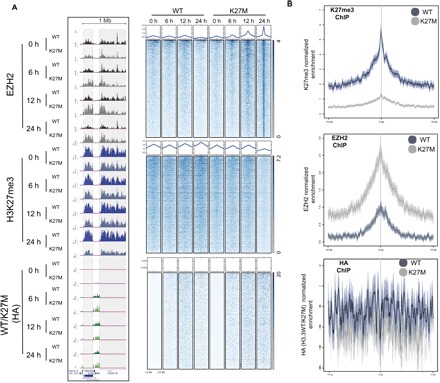
A methionine substitution at lysine-27 on histone H3 variants (H3K27M) characterizes ~80% of diffuse intrinsic pontine gliomas (DIPG) and inhibits polycomb repressive complex 2 (PRC2) in a dominant-negative fashion. Yet, the mechanisms for this inhibition and abnormal epigenomic landscape have not been resolved. Using quantitative proteomics, we discovered that robust PRC2 inhibition requires levels of H3K27M greatly exceeding those of PRC2, seen in DIPG. While PRC2 inhibition requires interaction with H3K27M, we found that this interaction on chromatin is transient, with PRC2 largely being released from H3K27M. Unexpectedly, inhibition persisted even after PRC2 dissociated from H3K27M-containing chromatin, suggesting a lasting impact on PRC2. Furthermore, allosterically activated PRC2 is particularly sensitive to H3K27M, leading to the failure to spread H3K27me from PRC2 recruitment sites and consequently abrogating PRC2's ability to establish H3K27me2-3 repressive chromatin domains. In turn, levels of polycomb antagonists such as H3K36me2 are elevated, suggesting a more global, downstream effect on the epigenome. Together, these findings reveal the conditions required for H3K27M-mediated PRC2 inhibition and reconcile seemingly paradoxical effects of H3K27M on PRC2 recruitment and activity.


Figures

Fig. 1
Fig. 1. PRC2 inhibition as a function of the H3K27M-to-PRC2 ratio.
(A) Samples are ranked left to right based on levels of H3K27me2-3, as detected by mass spectrometry (MS). mESC, mouse embryonic stem cell (n = 2); ASTRO, human astrocyte (n = 1); HEK 293, human embryonic kidney 293 cells (n = 2); WT GLIOMA, H3K27WT cortical glioma (n = 4); WT DIPG, H3K27WT DIPG (n = 2); NEURAL STEM, human neural stem cells (n = 2); mNEURON, mouse motor neuron (n = 3); K27M DIPG, DIPG with H3K27M (n = 2 for H3.1K27M and n = 2 for H3.3K27M); SUZ12-MPNST, malignant peripheral nerve sheath tumor that is SUZ12 null (n = 1). (B) PRC2 molecules per cell (average of EED, EZH2, and SUZ12) were determined by quantitative MS and are presented in the table, along with the relative ratio of H3K27M to PRC2. Levels of H3K27me2-3 determined by MS are presented in the chart below. DIPG and 293 T-REx cells with a large excess of K27M to PRC2 showed the most robust attenuation of H3K27me2-3 levels, while embryonic stem cells (mESC) with a more modest excess of K27M (~13-fold) showed a less robust loss in K27me2-3 relative to their WT counterparts (see table S1 for cell line details and fig. S1 for cell lines used in PRC2 quantitation analyses). (C) mESCs generated using CRISPR harboring either WT or a K27M mutation at H3F3A were differentiated to motor neurons (mNEURONs). Left: Sanger sequencing results for H3F3A K27M mESCs compared with WT mESCs. Right: Western blot validating the cell lines by H3K27M protein expression. (D) Left: Western blot validating the cell lines and increased K27M expression with significantly reduced levels of PRC2 core components in mNEURON. Islet1/2 served as an mNEURON marker. Right graph: Differentiation to mNEURON led to decreased K27me2-3 in K27M cells, relative to their mESC precursors, as measured through MS (n = 2 per cell type). (E) Top: Histone methyltransferase (HMT) assays containing PRC2 and increasing ratios of 8× oligonucleosomes comprising H3K27A or H3K27M and hemagglutinin (HA)–tagged H2A. Substrate oligonucleosomes were distinguishable by their reconstitution with H3-FLAG. Middle: Representative HMT assay shows levels of methylation and relative concentration of each HMT component. Bottom: Graphs quantitate the relative amount of 3H-SAM incorporated into histone H3-FLAG. Higher H3K27M-to-PRC2 ratios produce larger deficits in PRC2 activity (n = 3 per data point). Data plotted as means ± SD.
Fig. 2
Fig. 2. Abnormal recruitment and release of PRC2 from H3K27M-containing chromatin.
(A) 293 T-REx cells were induced to express C-terminal, FLAG-HA–tagged H3.3K27M (K27M) or FLAG-HA–tagged H3.3K27WT (WT) during the indicated time course. Dox, doxycycline. (B) Western blots revealed the progressive loss in H3K27me2-3 and gain of H3K36me2 as a function of time of H3.3K27M expression (note the slower migration of tagged H3.3 versions). GAPDH, glyceraldehyde-3-phosphate dehydrogenase. (C) Left: ChIP-seq for EZH2, H3K27me3, and HA-tagged WT/K27M histones performed at select time points and displayed as representative enrichment tracks. EZH2 peaks detected exclusively under the 6- and 12-hour conditions in K27M-expressing cells are highlighted in gray. Right: Heatmaps were generated by centering and rank ordering the 6-hour EZH2 peaks detected within EZH2 regions present only at the early 6- and 12-hour time points (1450 domains, 2325 peaks). Corresponding average density profiles are plotted at the top of the heatmaps. Peaks occurring specifically at the early time points (6- and 12-hour time points) strongly colocalize with K27M, are unique to the K27M cells, and are depleted by 24 hours.
Fig. 3
Fig. 3. PRC2 is recruited to normal target sites but is progressively less active on chromatin following H3K27M expression.
(A) ChIP-seq for EZH2, H3K27me3, and HA-tagged WT/K27M histones performed at select time points and displayed as representative enrichment tracks (left). PRC2 (EZH2) domains detected in both WT- and K27M-expressing cells across all time points are highlighted in gray. Right: Heatmaps centering on the 24-hour EZH2 peaks within the common PRC2 domains (616 domains, 2253 peaks) indicate strong EZH2 enrichment at that time point, decreased H3K27me3, and little detectable co-enrichment with H3K27M (HA). NTA, nitrilotriacetic acid. (B) To confirm the effects at the 24-hour time point, ChIP-seq was performed after 72 hours of H3.3K27M (K27M) and H3.3K27WT (WT) expression for EZH2, HA, and H3K27me3. Genome-wide enrichment plots centered on the common EZH2 domains corresponding to those in (A) are presented. Despite a loss in H3K27me3 in H3K27M cells relative to WT (top), EZH2 occupancy is increased, and the tagged histones are not enriched (middle and bottom, respectively). bp, base pair.
Fig. 4
Fig. 4. PRC2 purified from H3K27M cells or nucleosomes is less active.
(A) PRC2 containing 6×His-tagged EZH2 and FLAG-tagged EED was purified from 293 T-REx cells expressing H3.3K27M or H3.3K27WT for 3 days. Left: Western blot of levels of EZH2, EED, and histone H3 at each purification step. Note that histones and nonstoichiometric levels of EED or EZH2 were well removed through purification (see also fig. S4). IN, input; UB, unbound; B, bound. Lanes 8 and 16 (boxed) represent the final, purified PRC2 complex used in subsequent assays. Right: Silver staining showing the relative purity of each purified PRC2 complex. (B) Left: Purified PRC2 complexes as in (A) were subjected to HMT assays using 8× oligonucleosomes with H3-FLAG as substrate (300 nM) and increasing amounts (0.22, 0.66, 2, and 6 μM) of SAM [3H-SAM:SAM (1:9)]. Right: Quantification of the relative amounts of 3H-SAM incorporated into histone H3-FLAG substrate (n = 2). Data are plotted as means ± SD. (C) Coomassie blue staining shows recombinant PRC2 (EZH2, EED, SUZ12, and RBAP48) purified from SF9 cells. (D) Schematic representation of the method used to recover recombinant PRC2 after its association with different types of recombinant chromatin. Recombinant PRC2 (15 and 30 nM) was initially incubated with 8× oligonucleosomes containing HA-tagged H2A and either H3K27A or H3K27M (300 nM) for 1 hour at 30°C. PRC2 was then recovered by HA immunoprecipitation (IP), and the supernatant was collected. Equal amounts of unbound PRC2 (1× or 2×) was incubated with 3H-SAM (500 nM) using 8× oligonucleosomes comprising H3-FLAG as substrate (300 nM). (E) Left: A representative image of the HMT assays. Right: Quantitation of relative amount of 3H-SAM incorporated into histone H3-FLAG substrate (n = 3). Data are plotted as means ± SD.
Fig. 5
Fig. 5. H3K27M preferentially inhibits allosterically activated PRC2 in vitro and restricts remaining H3K27me3 in DIPG.
(A) Schematic representation of experiments in which PRC2 was incubated with 3H-SAM, increasing concentrations of H3K27me3 peptide, and 12× oligonucleosomes comprising HA-tagged H2A and either H3K27A or H3K27M. Oligonucleosomes containing FLAG-tagged H3 (300 nM) were added as a substrate. (B) One-half of the reaction was subject to autoradiography with subsequent quantification of the relative amount of 3H-SAM incorporated into the FLAG-tagged histone H3 (n = 3 per data point). Data are plotted as means ± SD. (C) The other half of the reaction was used to measure the amount of PRC2 retained on H3K27M- versus H3K27A-containing 12× nucleosomes by Western blot for EZH2 and EED after immunoprecipitation of the HA-tagged H2A-containing oligonucleosomes. The band intensity of EZH2 or EED served as a readout of EZH2 bound or EED bound on H3K27M- or H3K27A-containing 12× oligonucleosomes, as indicated (n = 3 per data point). Data are plotted as means ± SD. (D) Left: Representative H3K27me3 ChIP-seq tracks from patient-derived glioma cell lines with K27M (n = 2 cell lines) or WT H3 (n = 2 cell lines), showing very sharp, narrow peaks in the K27M glioma and much broader peaks in the WT glioma. Right: Genome-wide trend plot indicating that the remaining K27me3 peaks in the K27M glioma are more punctate in contrast to the WT glioma that show large, broad H3K27me3 signal indicative of typical polycomb domains.
Fig. 6
Fig. 6. Gain of K36me2 in H3K27M DIPG.
(A) H3K36me2 levels are significantly higher in H3K27M DIPG (n = 2 for H3.1K27M and n = 2 for H3.3K27M) than in WT DIPG/GLIOMA (n = 4 for H3K27WT cortical glioma and n = 2 for H3K27WT DIPG), as revealed by MS (see fig. S7). Data are plotted as means ± SD. (B) H3K36me2 ChIP-seq performed with glioma cell lines bearing either WT histone H3 (n = 2 total) or H3K27M, as indicated (n = 2; see table S1 for details). Left: The trend plot was generated by first identifying genes that displayed significant H3K36me2 enrichment above input for the H3K27M-bearing DIPG XIII cell line (1727 genes). Subsequently, average H3K36me2 levels were plotted for each glioma cell line across those genes (transcriptional start to end site). Right: As a control, H3K36me2 was plotted over all genes in the genome. Combined, H3K36me2-enriched genes in the H3K27M DIPG showed overall higher levels of H3K36me2 relative to glioma with WT histone H3. (C) Representative ChIP-seq tracks show generally elevated levels of H3K36me2 across these genes in H3K27M DIPG. Variations of the effect can be seen in these gliomas: Some genes are devoid of H3K36me2 in H3WT gain H3K36me2 in H3K27M, while others contain H3K36me2 in both H3WT and H3K27M glioma, yet the levels in the H3K27M cell lines are generally higher. TSS, transcriptional start site; TES, transcriptional end site.

References

    1. Talbert P. B., Henikoff S., Histone variants on the move: Substrates for chromatin dynamics. Nat. Rev. Mol. Cell Biol. 18, 115–126 (2017). - PubMed
    1. Jones C., Karajannis M. A., Jones D. T. W., Kieran M. W., Monje M., Baker S. J., Becher O. J., Cho Y.-J., Gupta N., Hawkins C., Hargrave D., Haas-Kogan D. A., Jabado N., Li X.-N., Mueller S., Nicolaides T., Packer R. J., Persson A. I., Phillips J. J., Simonds E. F., Stafford J. M., Tang Y., Pfister S. M., Weiss W. A., Pediatric high-grade glioma: Biologically and clinically in need of new thinking. Neuro Oncol. 19, 153–161 (2016). - PMC - PubMed
    1. Lewis P. W., Müller M. M., Koletsky M. S., Cordero F., Lin S., Banaszynski L. A., Garcia B. A., Muir T. W., Becher O. J., Allis C. D., Inhibition of PRC2 activity by a gain-of-function H3 mutation found in pediatric glioblastoma. Science 340, 857–861 (2013). - PMC - PubMed
    1. Bender S., Tang Y., Lindroth A. M., Hovestadt V., Jones D. T., Kool M., Zapatka M., Northcott P. A., Sturm D., Wang W., Radlwimmer B., Højfeldt J. W., Truffaux N., Castel D., Schubert S., Ryzhova M., Seker-Cin H., Gronych J., Johann P. D., Stark S., Meyer J., Milde T., Schuhmann M., Ebinger M., Monoranu C. M., Ponnuswami A., Chen S., Jones C., Witt O., Collins V. P., von Deimling A., Jabado N., Puget S., Grill J., Helin K., Korshunov A., Lichter P., Monje M., Plass C., Cho Y. J., Pfister S. M., Reduced H3K27me3 and DNA hypomethylation are major drivers of gene expression in K27M mutant pediatric high-grade gliomas. Cancer Cell 24, 660–672 (2013). - PubMed
    1. Mohammad F., Weissmann S., Leblanc B., Pandey D. P., Højfeldt J. W., Comet I., Zheng C., Johansen J. V., Rapin N., Porse B. T., Tvardovskiy A., Jensen O. N., Olaciregui N. G., Lavarino C., Suñol M., de Torres C., Mora J., Carcaboso A. M., Helin K., EZH2 is a potential therapeutic target for H3K27M-mutant pediatric gliomas. Nat. Med. 23, 483–492 (2017). - PubMed

Publication types

MeSH terms