

Emphysema quantification using chest CT: influence of radiation dose reduction and reconstruction technique
- PMID: 30402740
- PMCID: PMC6220000
- DOI: 10.1186/s41747-018-0064-3
Emphysema quantification using chest CT: influence of radiation dose reduction and reconstruction technique
Abstract
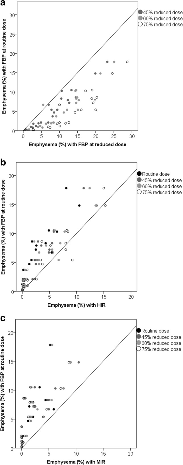
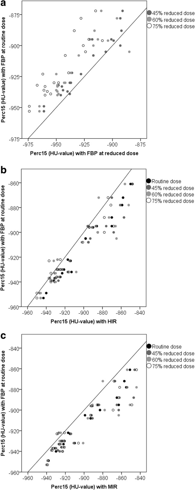
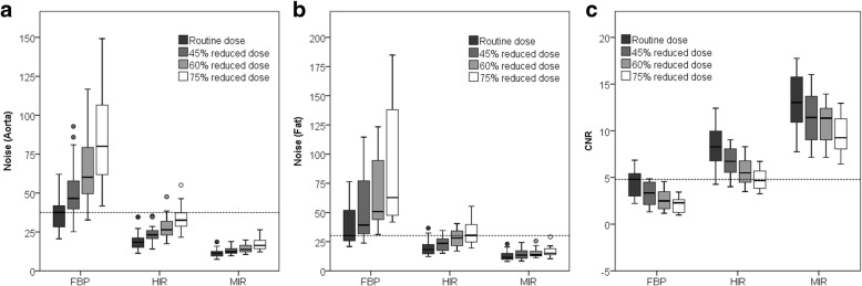
Background: Computed tomography (CT) emphysema quantification is affected by both radiation dose (i.e. image noise) and reconstruction technique. At reduced dose, filtered back projection (FBP) results in an overestimation of the amount of emphysema due to higher noise levels, while the use of iterative reconstruction (IR) can result in an underestimation due to reduced noise. The objective of this study was to determine the influence of dose reduction and hybrid IR (HIR) or model-based IR (MIR) on CT emphysema quantification.
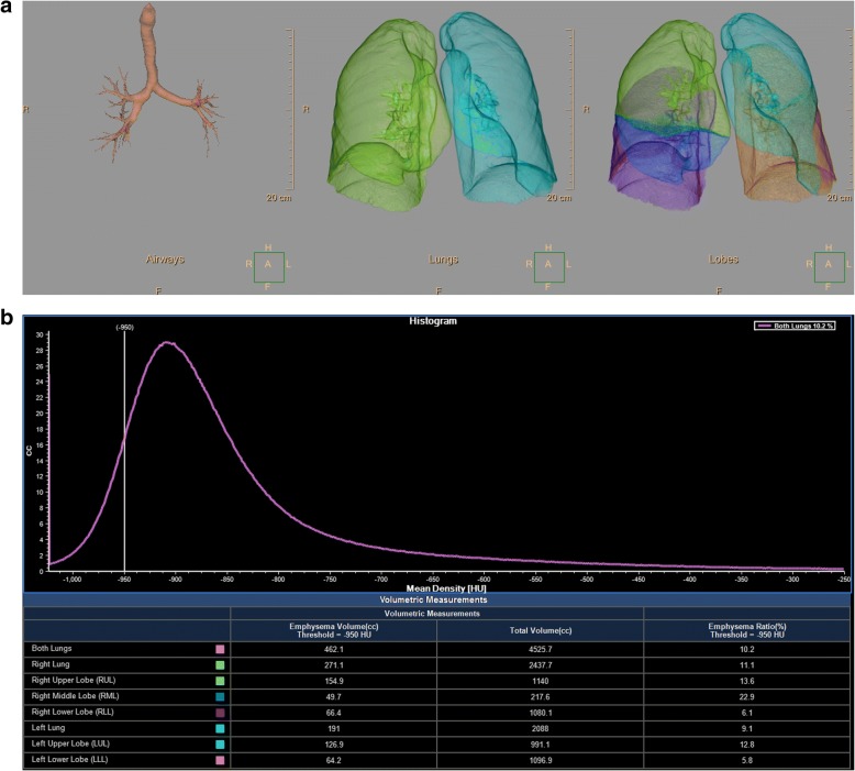
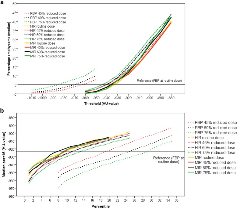
Methods: Twenty-two patients underwent inspiratory chest CT scan at routine radiation dose and at 45%, 60% and 75% reduced radiation dose. Acquisitions were reconstructed with FBP, HIR and MIR. Emphysema was quantified using the 15th percentile of the attenuation curve and the percentage of voxels below -950 HU. To determine whether the use of a different percentile or HU threshold is more accurate at reduced dose levels and with IR, additional measurements were performed using different percentiles and HU thresholds to determine the optimal combination.
Results: Dose reduction resulted in a significant overestimation of emphysema, while HIR and MIR resulted in an underestimation. Lower HU thresholds with FBP at reduced dose and higher HU thresholds with HIR and MIR resulted in emphysema percentages comparable to the reference. The 15th percentile quantification method showed similar results as the HU threshold method.
Conclusions: This within-patients study showed that CT emphysema quantification is significantly affected by dose reduction and IR. This can potentially be solved by adapting commonly used thresholds.
Keywords: Densitometry; Emphysema; Radiation dosage; Thorax; Tomography (x-ray computed).
Conflict of interest statement
Ethics approval and consent to participate
Institutional Review Board approval was obtained. Written informed consent was obtained from all subjects (patients) in this study.
Consent for publication
All authors provided consent for publication.
Competing interests
Julien Milles is an employee of Philips Healthcare. All other authors declare that they have no competing interests.
Publisher’s Note
Springer Nature remains neutral 12with regard to jurisdictional claims in published maps and institutional affiliations.
Figures
References
-
- Pompe Esther, Galbán Craig J., Ross Brian D., Koenderman Leo, ten Hacken Nick HT., Postma Dirkje S., van den Berge Maarten, de Jong Pim A., Lammers Jan-Willem J., Mohamed Hoesein Firdaus AA. Parametric response mapping on chest computed tomography associates with clinical and functional parameters in chronic obstructive pulmonary disease. Respiratory Medicine. 2017;123:48–55. doi: 10.1016/j.rmed.2016.11.021. - DOI - PMC - PubMed
-
- Madani Afarine, Van Muylem Alain, de Maertelaer Viviane, Zanen Jacqueline, Gevenois Pierre Alain. Pulmonary Emphysema: Size Distribution of Emphysematous Spaces on Multidetector CT Images—Comparison with Macroscopic and Microscopic Morphometry. Radiology. 2008;248(3):1036–1041. doi: 10.1148/radiol.2483071434. - DOI - PubMed
LinkOut - more resources
Full Text Sources
Miscellaneous