Review
. 2020 Jan;51(1):139-165.
doi: 10.1111/ejn.14254. Epub 2018 Dec 8.

Periodicity, repression, and the molecular architecture of the mammalian circadian clock

Affiliations
Review

Periodicity, repression, and the molecular architecture of the mammalian circadian clock

Clark Rosensweig et al. Eur J Neurosci. 2020 Jan.

Abstract

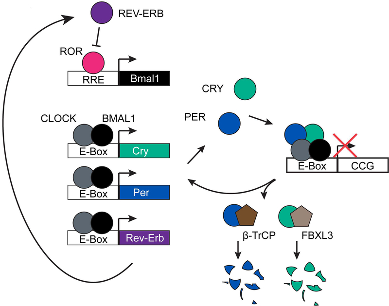
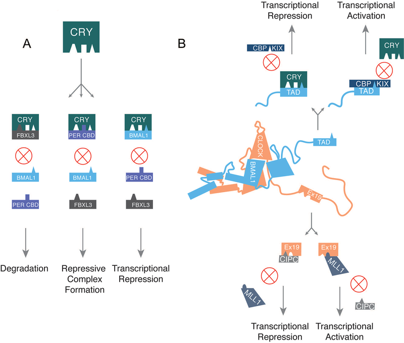
Large molecular machines regulate daily cycles of transcriptional activity and help generate rhythmic behavior. In recent years, structural and biochemical analyses have elucidated a number of principles guiding the interactions of proteins that form the basis of circadian timing. In its simplest form, the circadian clock is composed of a transcription/translation feedback loop. However, this description elides a complicated process of activator recruitment, chromatin decompaction, recruitment of coactivators, expression of repressors, formation of a repressive complex, repression of the activators, and ultimately degradation of the repressors and reinitiation of the cycle. Understanding the core principles underlying the clock requires careful examination of molecular and even atomic level details of these processes. Here, we review major structural and biochemical findings in circadian biology and make the argument that shared protein interfaces within the clockwork are critical for both the generation of rhythmicity and timing of the clock.

Keywords: clock; cryptochrome; period; rhythm; transcription.


Conflict of interest statement

Statement of Competing Financial Interests

All authors declare no competing financial or other interests.

Figures

Figure 1.
Figure 1.. Simple model of the mammalian transcription/translation feedback loop.
CLOCK and BMAL1 bind to E-boxes driving expression of their own repressors, CRYPTOCHROME and PERIOD. BMAL1 is also rhythmically expressed as a result of competitive binding at its promoter by the activator ROR and the repressor REV-ERB, which is under the control of CLOCK and BMAL1. Degradation of the repressors, CRY and PER, through interaction with FBXL3 and β–TrCP allows the cycle to begin again.
Figure 2.
Figure 2.. Molecular architecture of the activators CLOCK and BMAL1.
(A) On the top is the crystal structure of the CLOCK/BMAL1 heterodimer, encompassing the bHLH and PAS domains of each protein (PDB: 4F3L). CLOCK is shown in peach and BMAL1 is shown in blue. Below is a cartoon rendering of the heterodimer based on the structure, but with additional disordered regions drawn in. (B) Using available structural data, the structure of CLOCK is rendered in graphical form with domains of interest labeled and numbered based on the amino acid sequence of CLOCK from Mus musculus. (C) The structure of BMAL1 is rendered in graphical form with domains of interest labeled and numbered based on the amino acid sequence of BMAL1 from Mus musculus. (D) The PAS-A domains of CLOCK and BMAL1 have a reciprocal interaction in which the first α-helix of each PAS-A domain binds the β-sheet interface of its partner. (E) The PAS-B domains of CLOCK and BMAL1 interact through the β-sheet interface of BMAL1 and an α-helix of CLOCK, leaving a significant portion of CLOCK’s PAS-B available for other protein-protein interactions. (F) Residues identified as important for interaction between the CLOCK PAS-B domain, primarily its HI loop, and CRY are highlighted on the PAS-B structure in blue.
Figure 3.
Figure 3.. CRYPTOCHROME domain architecture.
(A) On the left, the CRY1 structure (PDB: 5T5X) is colored to show the α/β domain (spearmint) and the α-helical domain (blue). On the right, two graphical renderings of the CRY structure based on the actual crystal structures of CRY1 at left. Important features of the protein are labeled, including two cavities with roles in protein-protein interactions and a superficial structural feature (CC helix) that functions as a shared interface for competitive protein-protein interactions. (B) The structure of CRY1/2 is rendered in graphical form with features of interest labeled and numbered based on the amino acid sequences of CRY1 and CRY2 from Mus musculus. The first set of numbers refer to CRY1’s sequence and the second set in any pair refers to CRY2. CRY1 and CRY2 share the same basic structure outside of the variable C-terminal tail. Outside of the cavities, which are covered in the main text, CRY is notable for several flexible loops (the serine loop, the phosphate loop, and the interface loop), which function to some extent in physical interactions with other proteins. The serine loop and interface loop are both involved in binding to PER2. Additionally, the serine loop, along with α4, α15, and α16 contribute residues to the surface of the secondary pocket that play a role in binding to CLOCK’s HI loop. (C) An alignment of the CC helix and tail of murine CRYs. The alignment was made using Clustal Omega and visualized using ESPript 3 (Robert and Gouet, 2014). Although the CC helix is nearly identical, the tails of each CRY are highly divergent.
Figure 4.
Figure 4.. Divergence between CRY1 and CRY2.
(A) Two views of the CRY2 PHR structure (PDB: 4I6E) with all of the residues diverging between CRY1 and CRY2 labeled in blue. The vast majority of divergence is in one particular region of the α/β domain shown on the right. (B) The seven divergent residues at the secondary pocket are shown in blue on a surface representation of CRY2 (PDB: 4I6E).
Figure 5.
Figure 5.. PERIOD domain architecture.
(A) The PAS domain homodimer structures of PER1, PER2, and PER3 respectively. Overall structures are very similar. Each protein homodimerizes in an orthogonal orientation. The PAS-A and PAS-B domains of one PER1 subunit are circled in the figure on the left. N and C termini are labeled for each monomer of each PER complex.
(B) Using available structural data, the structure of PER2 is rendered in graphical form with domains of interest labeled and numbered based on the amino acid sequence of PER2 from Mus musculus. The region between the PAS-B domain and the CRY-Binding Domain (CBD) is shown as a flexible region, which reflects the fact that little structural information is available within this region. Within this region is the loosely defined Casein Kinase-Binding Domain (CKBD) and the even more loosely defined Proline-Rich Domain (PRD). No major roles have been ascribed to the PRD, but the CKBD contains several serines that are phosphorylated by casein kinase 1δ/ε. These serines (labeled here) are distal sites for regulation of PER stability. S478 is recognized by β-TrCP to target PER for degradation. S659, S662, S665, S668, and S671 are serially phosphorylated by CK1δ/ε and stabilize PER. Substitution of a glycine (S659G) at a homologous site in human PER2 is a known cause of Familial Advanced Sleep Phase Syndrome (FASPS) (Toh et al., 2001). CRY binds to PER at the distal C-terminal end of the protein beyond the PRD. PER1 and PER2 share a similar domain architecture, but PER3 diverges beyond the PAS domains and lacks a CBD altogether. Finally, three nuclear export sequences (NES) and a bipartite nuclear localization sequence (NLS) have been identified and validated in PER2 (Yagita et al., 2002).
Figure 6.
Figure 6.. Competitive protein-protein interactions drive clock function.
(A) CRY’s CC helix is involved in three mutually exclusive protein-protein interactions with FBXL3, PER, and BMAL1. The competitive interaction is depicted here along with the likely outcome of any given interaction. (B) Competitive interactions at two particular interfaces are potential drivers of oscillatory gene expression. The transcriptional repressor CRY competes with the transcriptional coactivator CBP to bind BMAL1’s TAD. As CRY levels build up throughout the activation phase of the clock, CRY supplants CBP at this interface causing a switch to a more repressive phase of the cycle. Similarly, MLL1 and CIPC both bind to CLOCK’s exon 19 region, likely in a mutually exclusive way. Interaction with MLL1 is critical for transcriptional activation at clock-regulated genes due to its role in chromatin decompaction. CIPC (and possibly PER proteins) functions as a repressor in part due to its ability to bind and sequester the exon 19 region of CLOCK. When the interaction partner at BMAL1’s TAD or CLOCK’s exon 19 switches, it allows for a transition in the phase of gene expression within the clock cycle.

Similar articles

Cited by

References

    1. Aguilar-Arnal L, Hakim O, Patel VR, Baldi P, Hager GL, and Sassone-Corsi P (2013). Cycles in spatial and temporal chromosomal organization driven by the circadian clock. Nature structural & molecular biology 20, 1206–1213. - PMC - PubMed
    1. Aguilar-Arnal L, Katada S, Orozco-Solis R, and Sassone-Corsi P (2015). NAD(+)-SIRT1 control of H3K4 trimethylation through circadian deacetylation of MLL1. Nature structural & molecular biology 22, 312–318. - PMC - PubMed
    1. Akashi M, Okamoto A, Tsuchiya Y, Todo T, Nishida E, and Node K (2014). A positive role for PERIOD in mammalian circadian gene expression. Cell reports 7, 1056–1064. - PubMed
    1. Akashi M, Tsuchiya Y, Yoshino T, and Nishida E (2002). Control of intracellular dynamics of mammalian period proteins by casein kinase I epsilon (CKIepsilon) and CKIdelta in cultured cells. Molecular and cellular biology 22, 1693–1703. - PMC - PubMed
    1. Albrecht U, Sun ZS, Eichele G, and Lee CC (1997). A differential response of two putative mammalian circadian regulators, mper1 and mper2, to light. Cell 91, 1055–1064. - PubMed

Publication types