

Unsolved Questions on the Anatomy of the Ventricular Conduction System
- PMID: 30403014
- PMCID: PMC6221866
- DOI: 10.4070/kcj.2018.0335
Unsolved Questions on the Anatomy of the Ventricular Conduction System
Abstract
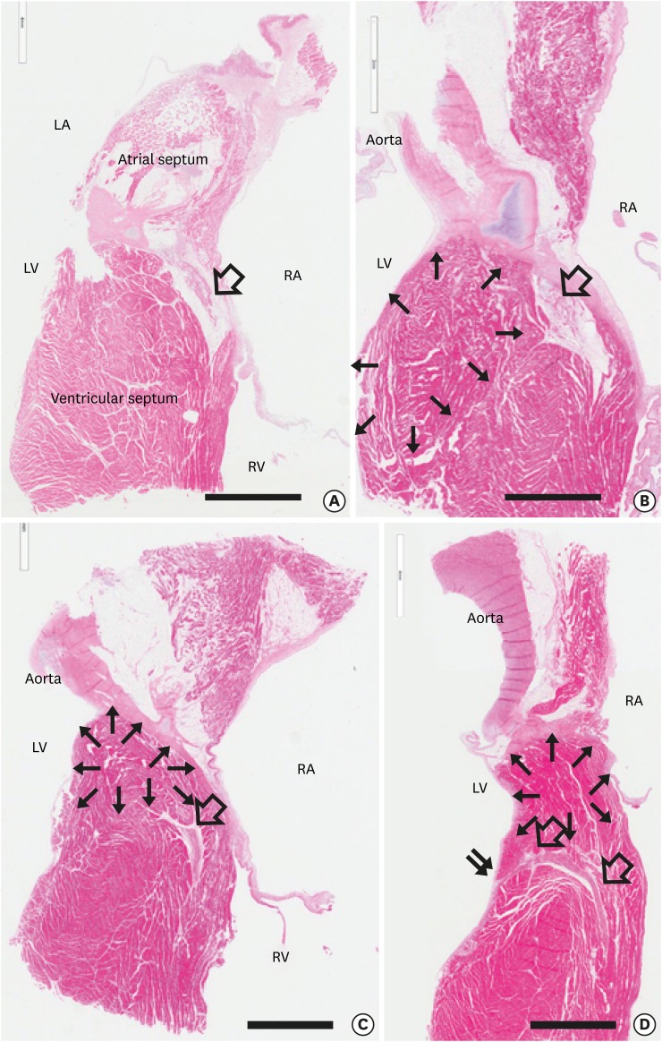
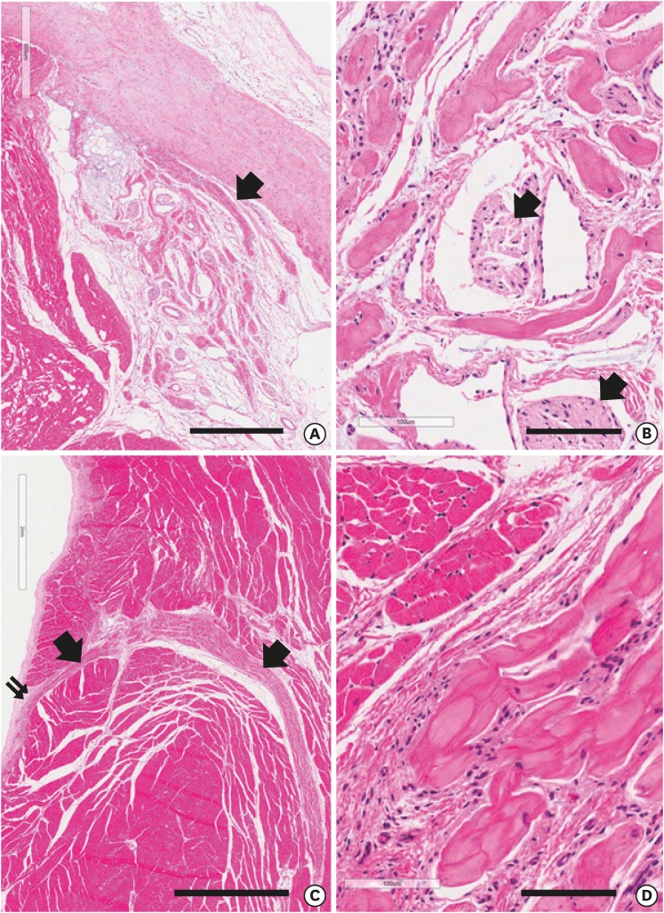
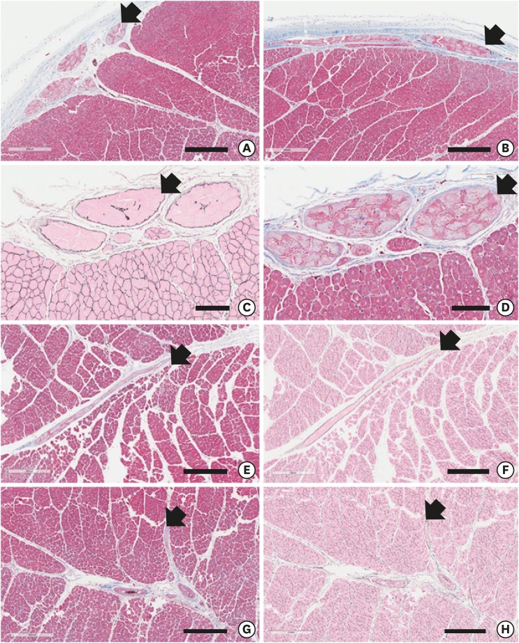
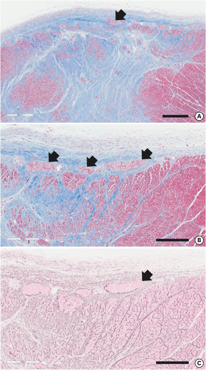
We reviewed the anatomical characteristics of the conduction system in the ventricles of human and ungulate hearts and then raised some questions to be answered by clinical and anatomical studies in the future. The ventricular conduction system is a 3-dimensional structure as compared to the 2-dimensional character of the atrial conduction system. The proximal part consisting of the atrioventricular node, the bundle of His and fascicles are groups of conducting cells surrounded by fibrous connective tissue so as to insulate from the underlying myocardium. Their location and morphological characters are well established. The bundle of His is a cord like structure but the left and right fascicles are broad at the proximal and branching at the distal part. The more distal part of fascicles and Purkinje system are linear networks of conducting cells at the immediate subendocardium but the intra-mural network is detected at the inner half of the ventricular wall. The papillary muscle also harbors Purkinje system not in the deeper part. It is hard to recognize histologically in human hearts but conducting cells as well as Purkinje cells are easily recognized in ungulate hearts. Further observation on human and ungulate hearts with myocardial infarct, we could find preserved Purkinje system at the subendocardium in contrast to the damaged system at the deeper myocardium. Further studies are necessary on the anatomical characteristics of this peripheral conduction system so as to correlate the clinical data on hearts with ventricular arrhythmias.
Keywords: Heart conduction system; Myocardial infarction; Purkinje fibers; Tachycardia, ventricular.
Copyright © 2018. The Korean Society of Cardiology.
Conflict of interest statement
The authors have no financial conflicts of interest.
Figures
Similar articles
-
Intramural Purkinje cell network of sheep ventricles as the terminal pathway of conduction system.Anat Rec (Hoboken). 2009 Jan;292(1):12-22. doi: 10.1002/ar.20827. Anat Rec (Hoboken). 2009. PMID: 19051253
-
Molecular characterization of the ventricular conduction system in the developing mouse heart: topographical correlation in normal and congenitally malformed hearts.Cardiovasc Res. 2001 Feb 1;49(2):417-29. doi: 10.1016/s0008-6363(00)00252-2. Cardiovasc Res. 2001. PMID: 11164852
-
Morphological study of the atrioventricular conduction system and Purkinje fibers in yak.J Morphol. 2017 Jul;278(7):975-986. doi: 10.1002/jmor.20691. Epub 2017 Apr 26. J Morphol. 2017. PMID: 28444887
-
The normal variants in the left bundle branch system.J Electrocardiol. 2017 Jul-Aug;50(4):389-399. doi: 10.1016/j.jelectrocard.2017.03.004. Epub 2017 Mar 14. J Electrocardiol. 2017. PMID: 28341304 Review.
-
[Anatomy of cardiac nodes and atrioventricular specialized conduction system].Rev Esp Cardiol. 2003 Nov;56(11):1085-92. doi: 10.1016/s0300-8932(03)77019-5. Rev Esp Cardiol. 2003. PMID: 14622540 Review. Spanish.
Cited by
-
Correlation between septal midwall late gadolinium enhancement on CMR and conduction delay on ECG in patients with nonischemic dilated cardiomyopathy.Int J Cardiol Heart Vasc. 2020 Jan 25;26:100474. doi: 10.1016/j.ijcha.2020.100474. eCollection 2020 Feb. Int J Cardiol Heart Vasc. 2020. PMID: 32021905 Free PMC article.
-
New Insights into the Development and Morphogenesis of the Cardiac Purkinje Fiber Network: Linking Architecture and Function.J Cardiovasc Dev Dis. 2021 Aug 7;8(8):95. doi: 10.3390/jcdd8080095. J Cardiovasc Dev Dis. 2021. PMID: 34436237 Free PMC article. Review.
-
Association between obesity and cardiac conduction defects.Front Cardiovasc Med. 2025 Jul 9;12:1476935. doi: 10.3389/fcvm.2025.1476935. eCollection 2025. Front Cardiovasc Med. 2025. PMID: 40703631 Free PMC article.
-
Remodeling of the Purkinje Network in Congestive Heart Failure in the Rabbit.Circ Heart Fail. 2021 Jul;14(7):e007505. doi: 10.1161/CIRCHEARTFAILURE.120.007505. Epub 2021 Jun 30. Circ Heart Fail. 2021. PMID: 34190577 Free PMC article.
-
Layer-specific speckle tracking analysis of left ventricular systolic function and synchrony in maintenance hemodialysis patients.BMC Cardiovasc Disord. 2020 Jan 9;20(1):126. doi: 10.1186/s12872-019-01324-z. BMC Cardiovasc Disord. 2020. PMID: 32160879 Free PMC article.
References
-
- Davies MJ. Pathology of conducting tissue of the heart. London: Butterworths; 1971.
-
- Seo JW, Chi JG, Lee SK. Cardiac conduction system-Morphological observations on the autopsy cases. Korean J Pathol. 1983;17:127–137.
-
- Ansari A, Ho SY, Anderson RH. Distribution of the Purkinje fibres in the sheep heart. Anat Rec. 1999;254:92–97. - PubMed
-
- Saffitz JE, Corradi D. The electrical heart: 25 years of discovery in cardiac electrophysiology, arrhythmias and sudden death. Cardiovasc Pathol. 2016;25:149–157. - PubMed
Publication types
LinkOut - more resources
Full Text Sources
