. 2018 Nov 7:7:e39866.
doi: 10.7554/eLife.39866.

Light-dependent pathways for dopaminergic amacrine cell development and function

Affiliations

Light-dependent pathways for dopaminergic amacrine cell development and function

Teona Munteanu et al. Elife. .

Abstract

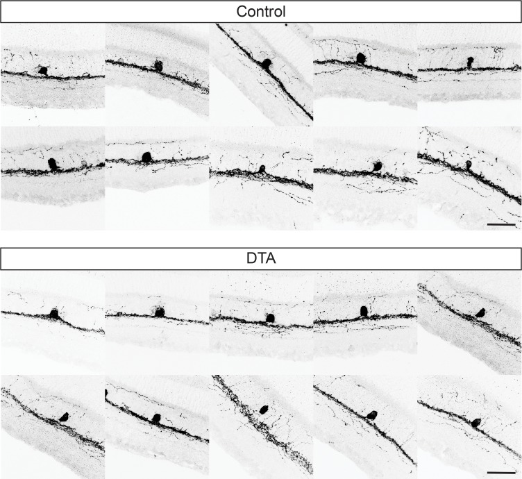
Retinal dopamine is a critical modulator of high acuity, light-adapted vision and photoreceptor coupling in the retina. Dopaminergic amacrine cells (DACs) serve as the sole source of retinal dopamine, and dopamine release in the retina follows a circadian rhythm and is modulated by light exposure. However, the retinal circuits through which light influences the development and function of DACs are still unknown. Intrinsically photosensitive retinal ganglion cells (ipRGCs) have emerged as a prime target for influencing retinal dopamine levels because they costratify with DACs in the inner plexiform layer and signal to them in a retrograde manner. Surprisingly, using genetic mouse models lacking specific phototransduction pathways, we find that while light influences the total number of DACs and retinal dopamine levels, this effect does not require ipRGCs. Instead, we find that the rod pathway is a critical modulator of both DAC number and retinal dopamine levels.

Keywords: development; dopamine; ipRGC; melanopsin; mouse; neuroscience; retina; rods.


Conflict of interest statement

TM, KN, AL, SP, JL, TS No competing interests declared

Figures

Figure 1.
Figure 1.. Light exposure during early retinal development is required to set TH-positive cell number and dopamine levels.
(A–B) TH+ cell number in animals bred and reared in either LD (yellow circles) or DD (black circles) from conception at P14 (n = 6 LD, n = 15 DD) (A) or Adult (n = 6 LD, n = 6 DD) (B) stages. Adult TH+ cell number could not be rescued by moving animals from DD to LD at P14 (hatched yellow circles, n = 4) (C) DA levels in adult retinas from animals reared in LD (yellow circles, n = 4) or DD (black circles, n = 4). (D) TH+ cells in whole mount WT adult retina (E–F) TH+ cell number (n = 9 Control, n = 4 TKO) (E) and DA levels (n = 6 Control, n = 6 TKO) (F) in Control (black circles) and TKO (open circles) adult retinas. *p < 0.05. Bars on plots represent mean.
Figure 2.
Figure 2.. Rod signaling influences TH-positive cell number and dopamine levels through ipRGC-independent pathways.
(A) Diagram depicting potential pathways by which light information could reach DACs (1. Via melanopsin signals relayed by ipRGCs, (2. Via rod signals relayed to DACs, (3. Via cone signals relayed to DACs, or (4. Via rod, cone, and/or melanopsin signals through ipRGC-dependent pathways. Dashed arrows represent indirect influence through multicellular circuits while solid arrows represent direct synaptic connectivity between subtypes. (B) Diagram depicting signaling pathways disrupted in MKO, RKO, CKO, and DTA mouse lines. Dashed arrows represent indirect influence through multicellular circuits while solid arrows represent direct synaptic connectivity between subtypes. (C–D) TH+ cell number and DA levels in Control (black circles, n = 9), MKO (blue circles, n = 6), RKO (red circles, n = 8), and CKO (green circles, n = 9) retinas from adult littermates. (E–F) TH+ cell number (n = 8 Control, n = 6 DTA) and DA levels (n = 6 Control, n = 6 DTA) in Control (black circles) and DTA (gray circles) retinas from adult littermates. (G) TH+ cell anatomy in WT and DTA adult retinal sections. We observed no morphological differences between TH+ cells in these two mouse lines. DA: dopamine, DAC: dopaminergic amacrine cell, MKO: animals lacking melanopsin phototransduction, RKO: animals lacking rod signaling, CKO: animals lacking cone signaling, DTA: animals where ipRGCs are ablated through expression of diphtheria toxin. Scale bar in (G) is 50 μm. *p < 0.05, bars on plots represent mean.
Figure 2—figure supplement 1.
Figure 2—figure supplement 1.. Examples of TH-positive cell stratification in WT and DTA retinas.
TH+ cell stratification in WT (top panels) and DTA (bottom panels) retinas show no differences in morphology. Scale bar is 50 μm.
Figure 3.
Figure 3.. Summary schematic for light-dependent influences on TH-positive cell number and retinal dopamine levels.
Rods signaling serves to increase TH+ cell number and DA levels through ipRGC-independent pathways. Dashed arrows represent indirect influence through multicellular circuits while solid arrows represent direct synaptic connectivity between subtypes. Red represents rod pathway signals, green represents cone pathway signals, and blue represents signals arising from melanopsin phototransduction. Neither ipRGC relay of rod, cone, nor melanopsin signals nor the cone pathway influence TH+ cell number or retinal DA levels. The rod pathway serves to increase the number of TH+ cells and increase retinal DA levels. R: Rod, C: Cone, DAC: dopaminergic amacrine cell, DA: Dopamine.

Similar articles

Cited by

References

    1. Altimus CM, Güler AD, Alam NM, Arman AC, Prusky GT, Sampath AP, Hattar S. Rod photoreceptors drive circadian photoentrainment across a wide range of light intensities. Nature Neuroscience. 2010;13:1107–1112. doi: 10.1038/nn.2617. - DOI - PMC - PubMed
    1. Besharse JC, McMahon DG. The retina and other Light-sensitive ocular clocks. Journal of Biological Rhythms. 2016;31:223–243. doi: 10.1177/0748730416642657. - DOI - PMC - PubMed
    1. Brooks E, Patel D, Canal MM. Programming of mice circadian photic responses by postnatal light environment. PLoS ONE. 2014;9:e97160. doi: 10.1371/journal.pone.0097160. - DOI - PMC - PubMed
    1. Calvert PD, Krasnoperova NV, Lyubarsky AL, Isayama T, Nicoló M, Kosaras B, Wong G, Gannon KS, Margolskee RF, Sidman RL, Pugh EN, Makino CL, Lem J. Phototransduction in transgenic mice after targeted deletion of the rod transducin alpha -subunit. PNAS. 2000;97:13913–13918. doi: 10.1073/pnas.250478897. - DOI - PMC - PubMed
    1. Chang B, Dacey MS, Hawes NL, Hitchcock PF, Milam AH, Atmaca-Sonmez P, Nusinowitz S, Heckenlively JR. Cone Photoreceptor Function Loss-3, a Novel Mouse Model of Achromatopsia Due to a Mutation in Gnat2. Investigative Opthalmology & Visual Science. 2006;47:5017–5021. doi: 10.1167/iovs.05-1468. - DOI - PubMed

Publication types

MeSH terms