Review
. 2019 Feb 10:81:585-605.
doi: 10.1146/annurev-physiol-021317-121245. Epub 2018 Nov 7.

Steps in Mechanotransduction Pathways that Control Cell Morphology

Affiliations
Review

Steps in Mechanotransduction Pathways that Control Cell Morphology

Haguy Wolfenson et al. Annu Rev Physiol. .

Abstract

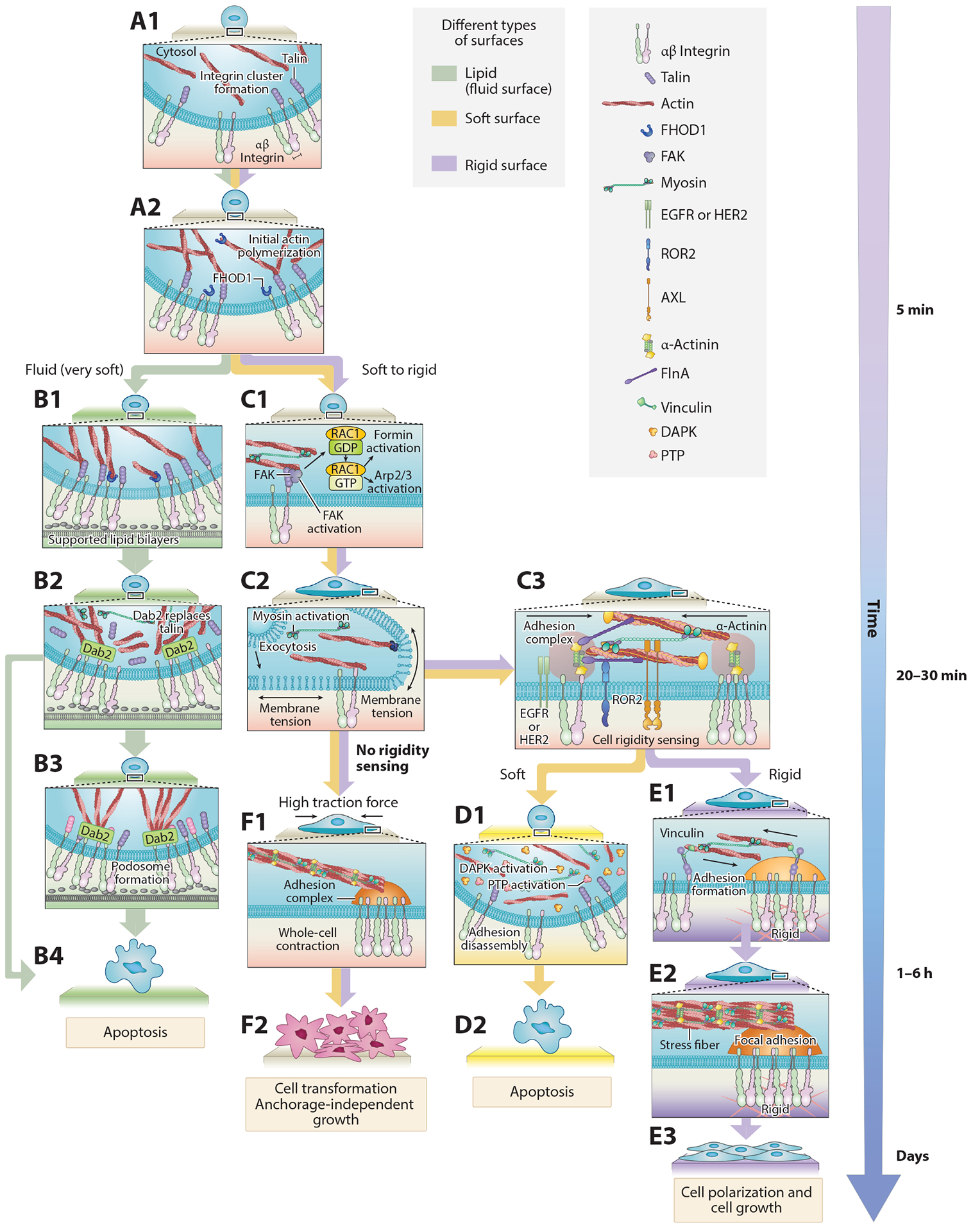
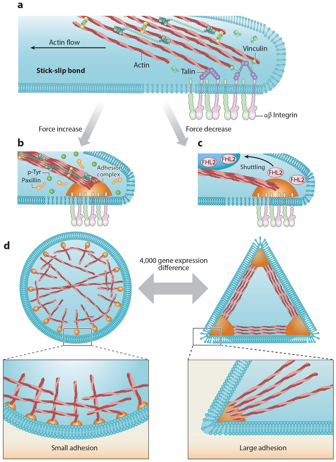
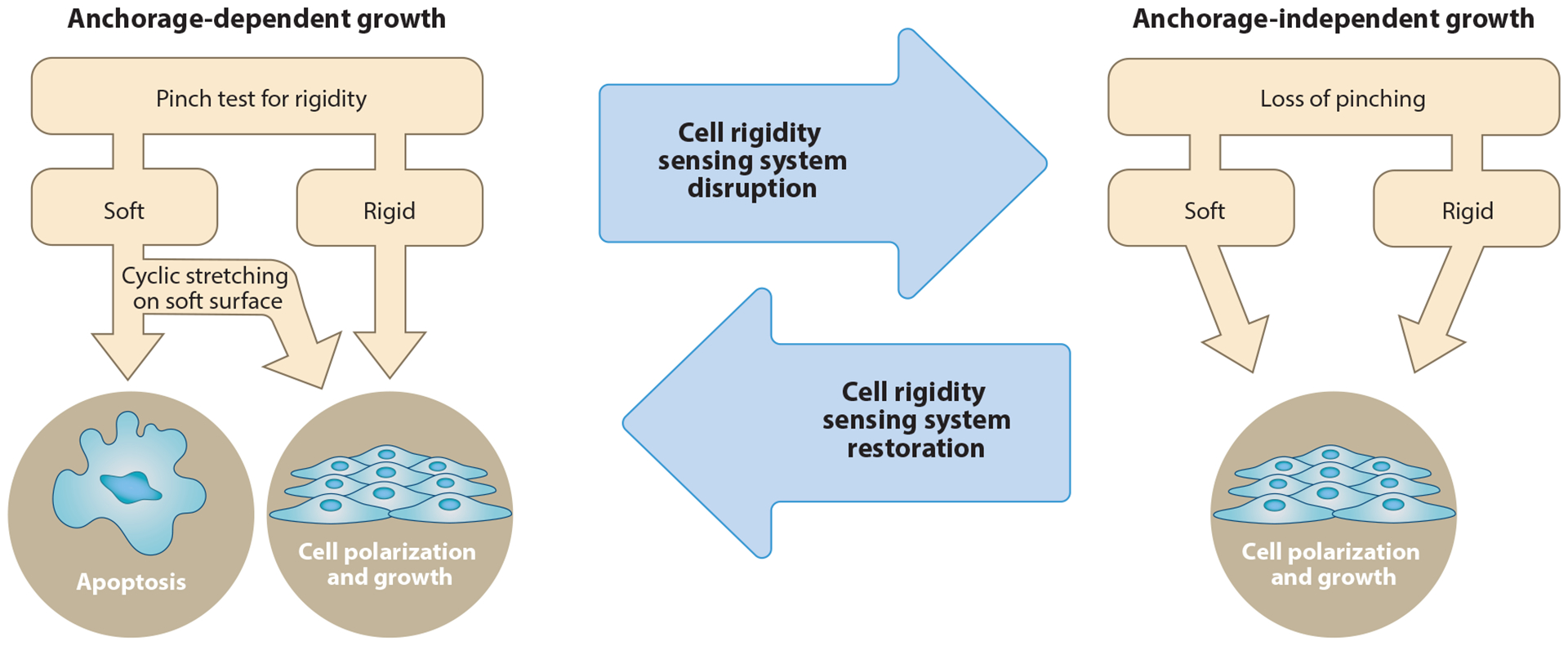
It is increasingly clear that mechanotransduction pathways play important roles in regulating fundamental cellular functions. Of the basic mechanical functions, the determination of cellular morphology is critical. Cells typically use many mechanosensitive steps and different cell states to achieve a polarized shape through repeated testing of the microenvironment. Indeed, morphology is determined by the microenvironment through periodic activation of motility, mechanotesting, and mechanoresponse functions by hormones, internal clocks, and receptor tyrosine kinases. Patterned substrates and controlled environments with defined rigidities limit the range of cell behavior and influence cell state decisions and are thus very useful for studying these steps. The recently defined rigidity sensing process provides a good example of how cells repeatedly test their microenvironment and is also linked to cancer. In general, aberrant extracellular matrix mechanosensing is associated with numerous conditions, including cardiovascular disease, aging, and fibrosis, that correlate with changes in tissue morphology and matrix composition. Hence, detailed descriptions of the steps involved in sensing and responding to the microenvironment are needed to better understand both the mechanisms of tissue homeostasis and the pathomechanisms of human disease.

Keywords: cell fate; cell morphology; cytoskeleton; integrin adhesions; mechanotransduction.


Figures

Figure 1
Figure 1
This diagram shows the basic cycles that underlie the shaping of cells and tissues. In most cases, intrinsic signals in cells activate motility processes, involving small G proteins, kinases, and actomyosin that change cell morphology or tug on neighboring cells to enable tests of the mechanical environment. The outcomes of those tests will cause cellular responses that can alter but often maintain the cell morphology. Although there is considerable variability in the duration of such cycles, the typical times are approximately 10 min for the complete cycle, meaning that many cycles will occur during a normal experimental period.
Figure 2
Figure 2
Mechanosensing occurs through a series of steps that involve adhesion formation, probing, and response. The cycle begins when (A1A2) the cell contacts RGD ligand and forms integrin clusters that activate actin polymerization (4, 44). (B1B4) Step-by-step mechanosensing of fibroblast cells on supported lipid bilayers (45, 114). (C1C3) When the cell edge encounters new matrix, it forms nascent integrin adhesions and begins to contract the matrix through contractile units in order to test its rigidity (32, 49, 54). (D1D2) Newly formed nascent adhesions disassemble when the matrix is soft. (E1E3) If the matrix is stiff enough and the forces reach a threshold of approximately 25 pN, adhesion is reinforced via recruitment of additional proteins from the cytoplasm. (F1F2) Transformed cells lose the rigidity sensing process and generate high traction forces on matrix. Abbreviations: AXL, anexelekto; Dab2, disabled homolog 2; DAPK, death-associated protein kinase; EGFR, epidermal growth factor receptor; FAK, focal adhesion kinase; FHOD1, formin homology (FH)1/FH2 domain-coating protein 1; FlnA, filamin A; HER2, human epidermal growth factor receptor 2; PTP, protein tyrosine phosphatase; RGD, arginylglycylaspartic acid; ROR2, receptor tyrosine kinase-like orphan receptor 2.
Figure 3
Figure 3
(a) Established adhesions experience drag forces from actin flowing to the cell center, resulting in a stick-slip behavior at the actin-adhesion interface (68). At later time points, when the adhesions mature, actin drag forces lead to repeated talin stretch-relaxation events that involve the binding of several vinculin molecules to a single talin (7). (b) Increasing forces cause increases in local tyrosine phosphorylation levels and reinforce adhesion formation. (c) Decreasing force leads to FHL2 shuttling to the nucleus (63). (d) Within 4 h of plating, cells on circular patterns show different expression levels of more than 4,000 genes compared with cells on triangle patterns (19). Abbreviations: FHL2, four and a half LIM domains protein 2; p-Tyr, phosphotyrosine.
Figure 4
Figure 4
Rigidity-sensing contractile units are needed for inhibition of growth on soft surfaces. When nontransformed cells properly form contractile units, they will detect when the matrix is stiff or soft and respond appropriately by growing or undergoing apoptosis, respectively. Depletion of many proteins in the contractile units causes transformation of the cells (32, 52, 99), which do not form contractile units and therefore cannot properly sense rigidity.
Figure 5
Figure 5
Involvement of adhesome components in cancer. Analysis is based on the Pathology Atlas, which is part of the Human Protein Atlas (https://www.proteinatlas.org). (a) Out of the 231 genes of the adhesome, 183 (~80%) are prognostic markers in cancer. Of those, 87 genes are unfavorable in specific cancer types (i.e., high mRNA expression correlates with shorter survival time), 46 genes are favorable in specific cancer types (high mRNA expression correlates with longer survival times), and 50 genes are favorable in some cancers and unfavorable in others. The remaining 48 genes are not prognostic, but 17 of those are still considered cancer related, i.e., often mutated in cancer. (b) Major categories of adhesome proteins involved in cancer and their division based on prognosis. Abbreviations: GAP, GTPase-activating proteins/GTPase-accelerating proteins; GEF, guanine nucleotide exchange factors.

References

    1. Taylor DA, Sampaio LC, Ferdous Z, Gobin AS, Taite LJ. 2018. Decellularized matrices in regenerative medicine. Acta Biomater. 74:74–89 - PubMed
    1. Yu Y, Alkhawaji A, Ding Y, Mei J. 2016. Decellularized scaffolds in regenerative medicine. Oncotarget 7:58671–83 - PMC - PubMed
    1. Dorland YL, Huveneers S. 2017. Cell–cell junctional mechanotransduction in endothelial remodeling. Cell. Mol. Life Sci 74:279–92 - PMC - PubMed
    1. Iskratsch T, Wolfenson H, Sheetz MP. 2014. Appreciating force and shape-the rise of mechanotransduction in cell biology. Nat. Rev. Mol. Cell Biol 15:825–33 - PMC - PubMed
    1. Roca-Cusachs P, Conte V, Trepat X. 2017. Quantifying forces in cell biology. Nat. Cell Biol 19:742–51 - PubMed

Publication types

LinkOut - more resources