. 2018 Nov 7;14(11):e1007753.
doi: 10.1371/journal.pgen.1007753. eCollection 2018 Nov.

Evidence that regulation of intramembrane proteolysis is mediated by substrate gating during sporulation in Bacillus subtilis

Affiliations

Evidence that regulation of intramembrane proteolysis is mediated by substrate gating during sporulation in Bacillus subtilis

Fernando H Ramírez-Guadiana et al. PLoS Genet. .

Abstract

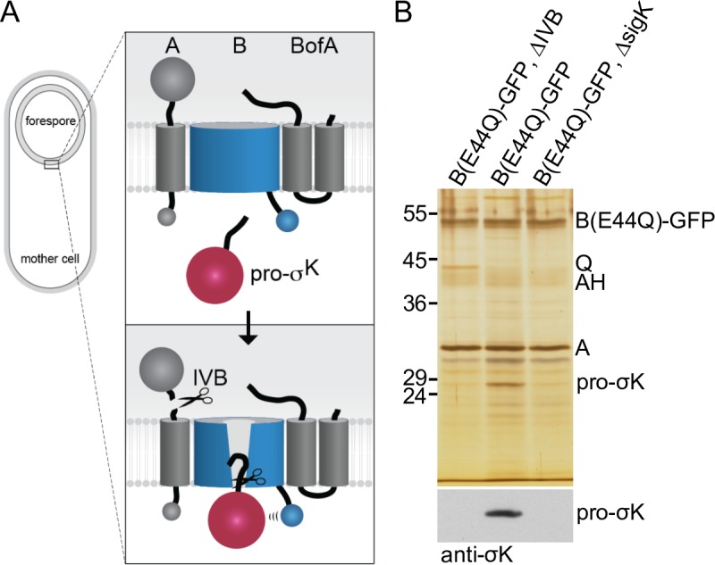
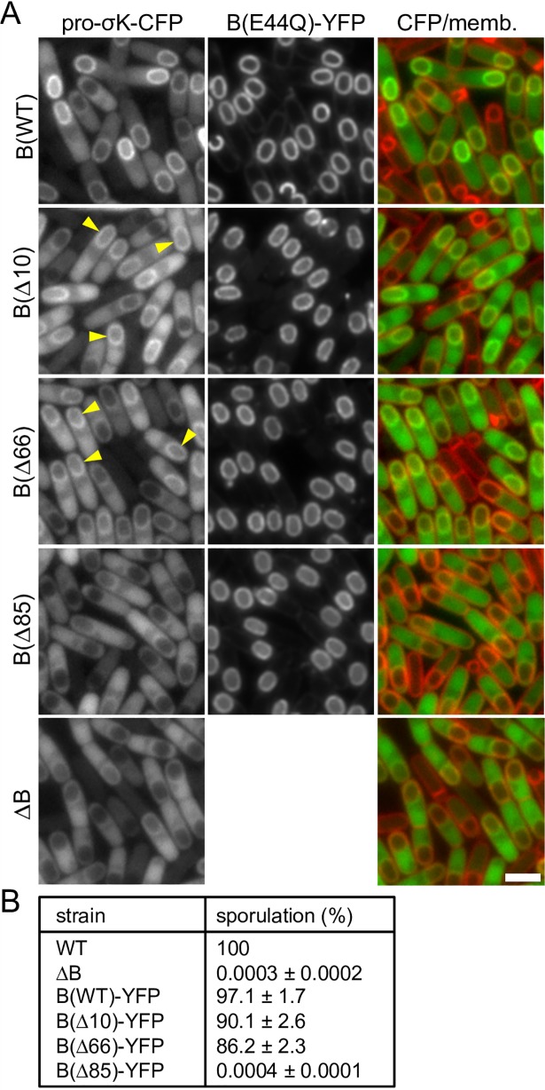
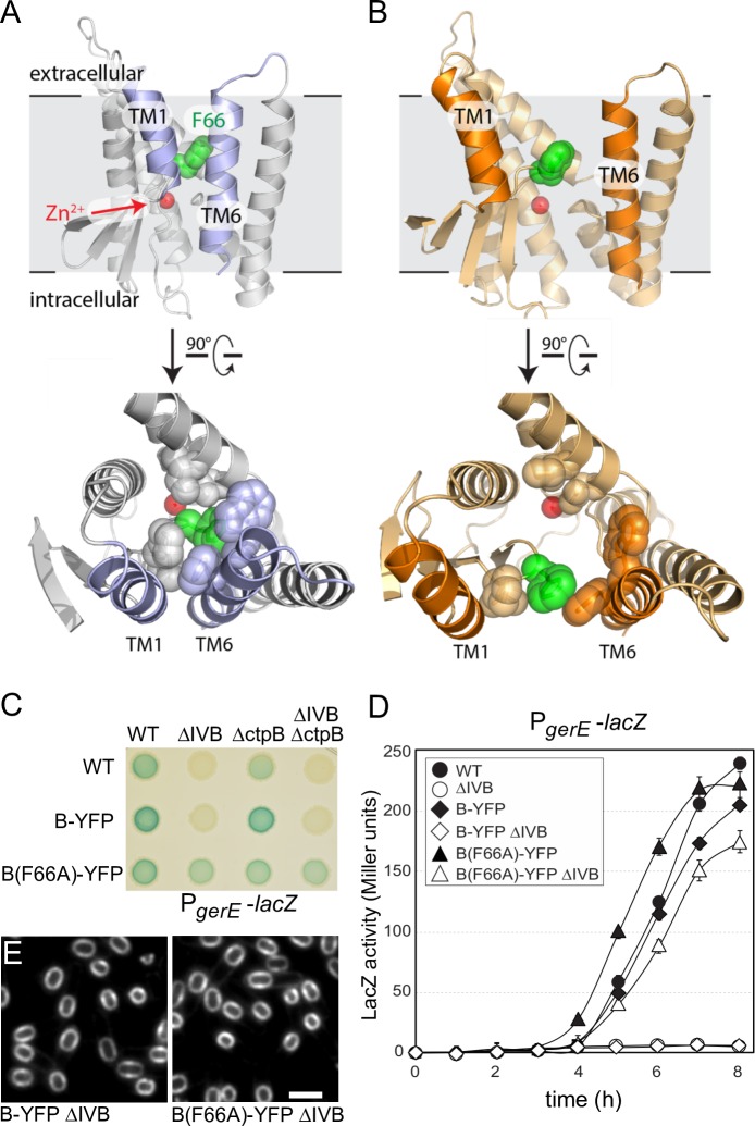
During the morphological process of sporulation in Bacillus subtilis two adjacent daughter cells (called the mother cell and forespore) follow different programs of gene expression that are linked to each other by signal transduction pathways. At a late stage in development, a signaling pathway emanating from the forespore triggers the proteolytic activation of the mother cell transcription factor σK. Cleavage of pro-σK to its mature and active form is catalyzed by the intramembrane cleaving metalloprotease SpoIVFB (B), a Site-2 Protease (S2P) family member. B is held inactive by two mother-cell membrane proteins SpoIVFA (A) and BofA. Activation of pro-σK processing requires a site-1 signaling protease SpoIVB (IVB) that is secreted from the forespore into the space between the two cells. IVB cleaves the extracellular domain of A but how this cleavage activates intramembrane proteolysis has remained unclear. Structural studies of the Methanocaldococcus jannaschii S2P homolog identified closed (substrate-occluded) and open (substrate-accessible) conformations of the protease, but the biological relevance of these conformations has not been established. Here, using co-immunoprecipitation and fluorescence microscopy, we show that stable association between the membrane-embedded protease and its substrate requires IVB signaling. We further show that the cytoplasmic cystathionine-β-synthase (CBS) domain of the B protease is not critical for this interaction or for pro-σK processing, suggesting the IVB-dependent interaction site is in the membrane protease domain. Finally, we provide evidence that the B protease domain adopts both open and closed conformations in vivo. Collectively, our data support a substrate-gating model in which IVB-dependent cleavage of A on one side of the membrane triggers a conformational change in the membrane-embedded protease from a closed to an open state allowing pro-σK access to the caged interior of the protease.


Conflict of interest statement

The authors have declared that no competing interests exist.

Figures

Fig 1
Fig 1. Pro-σK resides in a membrane complex with the B protease in a IVB-dependent manner.
(A) Schematic model of regulated intramembrane proteolysis during sporulation. Prior to forespore signaling, A and BofA hold B in a closed conformation in the membranes surrounding the forespore. IVB-dependent cleavage of A triggers a conformational change in B allowing pro-σK access to the catalytic center of the protease. Pro-σK processing then leads to late mother cell gene expression and spore maturation (not depicted in the diagram). For simplicity, A and BofA are shown interacting with B but not each other. Co-immunoprecipitation and cytological assays [33] suggest A and BofA interact in the absence of B. (B) Silver-stained gel of immunoprecipitated proteins from detergent-solubilized membrane preparations of the indicated strains at hour 4 of sporulation. Proteins indicated on the right were identified by mass spec or from immunoprecipitations using mutant strains. Immunoblot of the same samples using anti-σK antibodies is shown below.
Fig 2
Fig 2. Pro-σK-CFP localization to the membranes surrounding the forespore requires IVB protease activity.
Representative images of the indicated strains at hour 4 of sporulation. (A) Pro-σK-CFP co-localizes with B(E44Q)-YFP in the mother-cell membranes surrounding the forespore in cells harboring the IVB signaling protease. In the absence of IVB or a catalytic mutant (S378A) of IVB, pro-σK-CFP localizes to the mother-cell cytoplasm with some enrichment on the nucleoid. Membranes were stained with the fluorescent dye TMA-DPH. Larger fields of cells over a sporulation time course and quantification of forespore-associated pro-σK-CFP can be found in S3 and S4 Figs. (B) Pro-σK-CFP co-localizes with the mother-cell nucleoid in strains harboring the catalytically active B (site-2) protease and the IVB (site-1) signaling protease. In the absence of IVB or in the catalytic mutant IVB(S378A), pro-σK localizes in the mother-cell cytoplasm with some enrichment on the nucleoid. DNA was stained with DAPI. Images were scaled identically. Scale bar indicate 2 μm.
Fig 3
Fig 3. Pro-σK-CFP localization to the membranes surrounding the forespore requires the B protease.
Representative images of the indicated strains at hour 4 of sporulation. Pro-σK-CFP localizes to the membranes surrounding the forespore in cells harboring B(E44Q)-YFP, in the absence of A, BofA, or both. In sporulating cells lacking the B protease, pro-σK localizes to the mother-cell cytoplasm with some enrichment on the nucleoid. The cytoplasmic pro-σK-CFP signal is more pixelated in the strains lacking A and/or BofA compared to the ΔB mutant (see text). Images were scaled identically. Scale bar indicates 2 μm. Quantification of forespore-associated pro-σK-CFP and larger fields of cells for all 5 strains over a sporulation time course can be found in S4 and S5 Figs.
Fig 4
Fig 4. Pro-σK-CFP localizes to the membranes of vegetatively growing cells when B(E44Q) is co-expressed.
(A) Representative images of the indicated strains after 1.5 hours of induction. In the absence of the B protease, pro-σK-CFP localizes in the cytoplasm. In the presence of wild-type B-YFP, pro-σK-CFP gets processed and localizes to the nucleoid. Pro-σK-CFP co-localizes with B(E44Q)-YFP in the septal and cytoplasmic membranes when co-expressed during vegetative growth. Pro-σK-CFP remains cytoplasmic when wild-type B-YFP is co-expressed with its inhibitors A and BofA. The cytoplasmic Pro-σK-CFP signal is weaker and more pixelated in the strain expressing B(E44Q)-YFP compared to those lacking B or co-expressing A and BofA (see text). All images were scaled identically. Larger fields of cells over the induction time course can be found in S6 Fig. Scale bar indicates 2 μm. (B) Immunoblot of the same strains in (A) using anti-σK antibodies showing pro-σK-CFP processing in cells expressing B-YFP but not in cells expressing the E44Q mutant, the inhibitors A and BofA, or in a strain that does not express B during vegetative growth. σA was used to control for loading. Molecular weight markers (in kDa) are indicated to the left. An anti-GFP immunoblot to assess the relative amount of liberated (free) CFP in the four strains can be found in S2C Fig.
Fig 5
Fig 5. The CBS domain of the B protease is not critical for pro-σK-CFP localization or efficient sporulation.
(A) Representative images of the indicated strains at hour 4.5 of sporulation. Pro-σK-CFP localizes around the forespore in cells producing B(E44Q)-YFP or 10 and 66 amino acid C-terminal truncations of the catalytic mutant. Pro-σK-CFP localizes to the mother-cell cytoplasm with some enrichment on the nucleoid in the presence of a larger 85 amino acid truncation similar to sporulating cells lacking the B protease. Yellow carets highlight the localization of pro-σK-CFP around the forespore. All images were scaled identically. Quantification of forespore-associated pro-σK-CFP and larger fields of cells for all 5 strains over a sporulation time course can be found in S4 and S7 Figs. Scale bar indicates 2 μm. (B) Table of sporulation efficiencies of the indicated strains (n = 3). All strains have untagged pro-σK and the B protease fusions have intact catalytic residues.
Fig 6
Fig 6. Evidence that B adopts open and closed conformations in vivo.
Models of the closed (A) and open (B) conformations of the B protease domain based on the mjS2P structures. Phenylalanine 66 (green) in the membrane-reentrant β-loop that is predicted to occlude substrate and help stabilize the closed conformation and the catalytic Zn2+ ion (red ball) are indicated. Open and closed conformations rotated 90˚ are shown below. The residues in the catalytic core that are within 5 Å of F66 in the closed conformation are shown in both states. (C) B(F66A)-YFP activates σK in the absence of IVB and an auxiliary signaling protease CtpB. σK activity was monitored using a σK-responsive promoter (PgerE) fused to lacZ. Image of a sporulation agar plate containing X-gal with the indicated strains after 24 hours of incubation at 37˚C. (D) Liquid β-galactosidase assay during a sporulation time course (n = 3). Only B(F66A) activates σK in the absence of IVB (open triangles). Liquid β-galactosidase assays of additional strains and immunoblot analysis of pro-σK processing can be found in S11 Fig. (E) B(F66A)-YFP localizes to the membranes surrounding the forespore. Representative images of the indicated strains at hour 4.5 of sporulation. Scale bar indicates 2 μm. Additional controls can be found in S12 Fig.

Similar articles

Cited by

References

    1. Brown MS, Ye J, Rawson RB, Goldstein JL. Regulated intramembrane proteolysis: a control mechanism conserved from bacteria to humans. Cell. 2000;100(4):391–8. . - PubMed
    1. Sun L, Li X, Shi Y. Structural biology of intramembrane proteases: mechanistic insights from rhomboid and S2P to gamma-secretase. Curr Opin Struct Biol. 2016;37:97–107. 10.1016/j.sbi.2015.12.008 . - DOI - PubMed
    1. Wolfe MS, Kopan R. Intramembrane proteolysis: theme and variations. Science. 2004;305(5687):1119–23. 10.1126/science.1096187 . - DOI - PubMed
    1. Kroos L, Akiyama Y. Biochemical and structural insights into intramembrane metalloprotease mechanisms. Biochim Biophys Acta. 2013;1828(12):2873–85. 10.1016/j.bbamem.2013.03.032 ; PubMed Central PMCID: PMCPMC3793210. - DOI - PMC - PubMed
    1. Rawson RB. The site-2 protease. Biochim Biophys Acta. 2013;1828(12):2801–7. 10.1016/j.bbamem.2013.03.031 . - DOI - PubMed

Publication types

LinkOut - more resources