. 2018 Nov 7;13(11):e0205969.
doi: 10.1371/journal.pone.0205969. eCollection 2018.

DNMTs and SETDB1 function as co-repressors in MAX-mediated repression of germ cell-related genes in mouse embryonic stem cells

Affiliations

DNMTs and SETDB1 function as co-repressors in MAX-mediated repression of germ cell-related genes in mouse embryonic stem cells

Daiki Tatsumi et al. PLoS One. .

Abstract

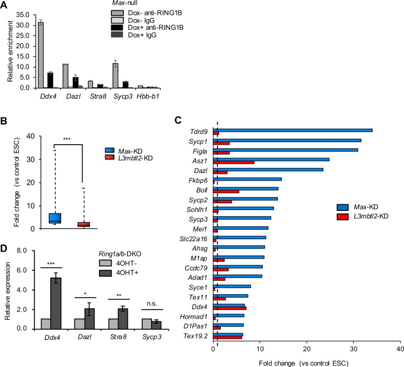
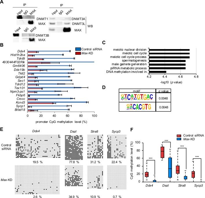
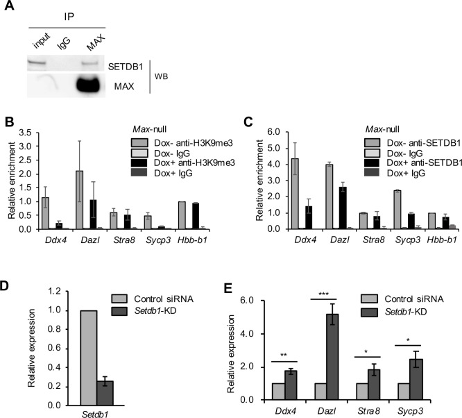
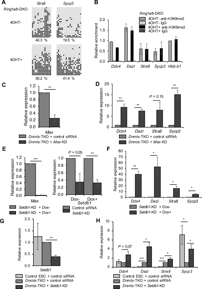
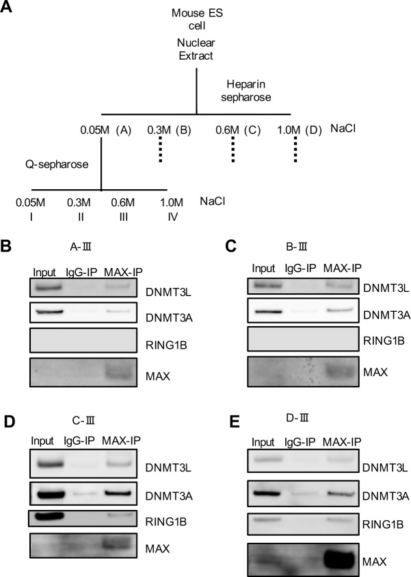
In embryonic stem cells (ESCs), the expression of development-related genes, including germ cell-related genes, is globally repressed. The transcription factor MAX represses germ cell-related gene expression in ESCs via PCGF6-polycomb repressive complex 1 (PRC1), which consists of several epigenetic factors. However, we predicted that MAX represses germ cell-related gene expression through several additional mechanisms because PCGF6-PRC1 regulates the expression of only a subset of genes repressed by MAX. Here, we report that MAX associated with DNA methyltransferases (DNMTs) and the histone methyltransferase SETDB1 cooperatively control germ cell-related gene expression in ESCs. Both DNA methylation and histone H3 lysine 9 tri-methylation of the promoter regions of several germ cell-related genes were not affected by knockout of the PRC1 components, indicating that the MAX-DNMT and MAX-SETDB1 pathways are independent of the PCGF6-PRC1 pathway. Our findings provide insights into our understanding of MAX-based repressive mechanisms of germ cell-related genes in ESCs.


Conflict of interest statement

IM is employed by and received salary from HaploPharma Inc., Chuo-ku, Tokyo, Japan. This does not alter our adherence to all the PLOS ONE policies on sharing data and materials.

Figures

Fig 1
Fig 1. Contribution of PCGF6-PRC1 to MAX-mediated gene repression.
(A) ChIP-qPCR analyses of Max-null ESCs (Dox+) and control ESCs (Dox−) using anti-RING1B antibody or control IgG in the promoter region of the late PGC markers and of hemoglobin β (Hbb-b1) as a negative control of MAX localization. Relative ratio of immunoprecipitated chromatin to input chromatin (% of input) was determined using real-time PCR. Data are presented as relative % of input normalized against % of input using anti-RING1B antibody in Hbb-b1 in control ESCs. The bar graph represents mean ± standard error of the mean (SEM) of two independent experiments. (B) Expression changes of 85 germ cell–related up-regulated genes (n = 3, > 2-fold change, one-way analysis of variance [ANOVA] P < 0.05) in Max-KD or L3mbtl2-KD ESCs compared with control ESCs based on our microarray data (GSE45181) [2]. Fold-change in the expression is shown by a box-whisker plot. The lines inside the boxes show the median. The whiskers indicate the minimum and maximum. ***P < 0.001 (Mann-Whitney U-test). (C) Fold-change in the expression of significantly up-regulated germ cell–related genes (top 22 of 85 germ cell-related upregulated genes in Fig 1B) in Max-KD and L3mbtl2-KD ESCs. The expression in control ESCs was set as 1.0 (dotted line). (D) Relative expression of the late PGC markers in Ring1a/b-DKO ESCs (4OHT+) determined by qRT-PCR. The expression in control ESCs (4OHT−) was set as 1.0. Values are plotted as mean ± SEM of 3 biological replicates. n.s.: not significant, *P < 0.05, **P < 0.01, ***P < 0.001 (Student’s t-test).
Fig 2
Fig 2. MAX-mediated repression of germ cell–related genes through DNA methylation.
(A) Samples immunoprecipitated using anti-MAX antibody or control IgG were analyzed by Western blotting using anti-DNMT antibodies. Principally, the same result was obtained in two independent experiments. The un-cropped data of these images are shown in S4A Fig. (B) Levels of CpG methylation of genes with a DMR in close proximity (±300 bp) for Max-KD ESCs and control ESCs from TMS. The ratios of methylated CpGs in the regions ±300 bp of the genes are shown. Values are plotted as mean ± SEM of 2 biological replicates. (C) GO analysis of 17 genes with DMRs hypomethylated in Max-KD ESCs compared with control ESCs. GO terms with corrected P value < 0.05 (top 7) are shown. (D) Motif analyses of 17 DMRs showed significant enrichment of E-box–like sequences. Motif sequences with the lowest q value (top 2) are shown. (E) DNA methylation status of the promoter regions of the late PGC markers in control and Max-KD ESCs, as determined by bisulfite sequencing. The filled and open circles indicate methylated- and un-methylated CpGs, respectively. The data shown were combined from two independent experiments. The percentage of methylated CpGs is indicated. (F) Box-whisker plots of the CpG methylation levels shown in Fig 2E. The lines inside the boxes show the median. The whiskers indicate the minimum and maximum. ***P < 0.001 (Mann-Whitney U-test).
Fig 3
Fig 3. MAX-mediated repression of germ cell–related genes through H3K9me3.
(A) Samples immunoprecipitated using anti-MAX antibody or control IgG were analyzed by Western blotting using anti-SETDB1 antibody. Principally, the same result was obtained in two independent experiments. The un-cropped data of this image is shown in S5B Fig. (B, C) ChIP-qPCR analyses of Max-null ESCs (Dox+) and control ESCs (Dox−) using anti-H3K9me3 antibody (B), anti-SETDB1 antibody (C) or control IgG. The data are displayed in the same way as in Fig 1A. (D) KD efficiency of Setdb1 in ESCs at day 2 post-siRNA treatment, as determined by RT-qPCR. (E) Relative expression of the late PGC markers in Setdb1-KD ESCs, as determined by qRT-PCR. The expression in control ESCs was set as 1.0. Values are plotted as the mean ± SEM of 3 biological replicates. *P < 0.05, **P < 0.01, ***P < 0.001 (Student’s t-test).
Fig 4
Fig 4. RING1A/B-independent DNA and H3K9 methylation, and a cooperative effect of Dnmts-TKO and Setdb1-KD on repression of the late PGC markers.
(A) DNA methylation status of the promoter regions of Stra8 and Sycp3 in control (4OHT−) and Ring1a/b-DKO ESCs (4OHT+) determined by bisulfite sequencing. The data are displayed in the same way as in Fig 2E. (B) ChIP-qPCR analyses of Ring1a/b-DKO ESCs (4OHT+) and control ESCs (4OHT−) using anti-H3K9me3 antibody or control IgG. The data are displayed in the same way as in Fig 1A. (C) KD efficiency of Max in Dnmts-TKO ESCs at day 3 post-siRNA treatment, as determined by RT-qPCR. (D) Relative expression of the late PGC markers in Max-KD/Dnmts-TKO ESCs, as determined by qRT-PCR. The expression in Dnmts-TKO ESCs with control siRNA treatment was set as 1.0. (E) KO and KD efficiency of Max (left panel) and Setdb1 (right panel) in Setdb1-KD/Max-null ESCs at day 3 post-siRNA treatment, as determined by RT-qPCR. KD efficiency of Setdb1 is normalized with the expression of Setdb1 in Max-null ESCs with control siRNA treatment. (F) Relative expression of the late PGC markers in Setdb1-KD/Max-KO ESCs, as determined by qRT-PCR. The expression in Setdb1-KD ESCs with Max expression (Dox-) was set as 1.0. (G) KD efficiency of Setdb1 in Dnmts-TKO ESCs at day 2 post-siRNA treatment, as determined by RT-qPCR. (H) Relative expression of the late PGC markers in Setdb1-KD/Dnmts-TKO ESCs, as determined by qRT-PCR. The expression in Dnmts-TKO ESCs with control siRNA treatment was set as 1.0. Values are plotted as the mean ± SEM of 3 biological replicates. *P < 0.05, **P < 0.01, ***P < 0.001 (Student’s t-test).
Fig 5
Fig 5. Fractionation of MAX-interacting complexes.
(A) Schematic illustration of the fractionation of ESC nuclear extracts by column chromatography. Nuclear extracts were prepared from VV3 ESCs and fractionated into fractions A-D on a heparin sepharose column. Fractions A-D were further fractionated into fractions I-IV on a Q sepharose column. (B to E) Samples immunoprecipitated using anti-MAX antibody or control IgG for fraction A-III (B), B-III (C), C-III (D), or D-III (E) were analyzed by Western blotting using anti-DNMT, anti-RING1B, or anti-MAX antibodies. Principally, the same result was obtained in two independent experiments. The un-cropped data of these images are shown in S6A–S6D Fig, respectively.

Similar articles

Cited by

References

    1. Orkin SH, Hochedlinger K. Chromatin connections to pluripotency and cellular reprogramming. Cell. 2011; 145: 835–850. 10.1016/j.cell.2011.05.019 - DOI - PMC - PubMed
    1. Maeda I, Okamura D, Tokitake Y, Ikeda M, Kawaguchi H, Mise N, et al. Max is a repressor of germ cell-related gene expression in mouse embryonic stem cells. Nat Commun. 2013; 4: 1754 10.1038/ncomms2780 - DOI - PubMed
    1. Suzuki A, Hirasaki M, Hishida T, Wu J, Okamura D, Ueda A, et al. Loss of MAX results in meiotic entry in mouse embryonic and germline stem cells. Nat Commun. 2016; 7: 11056 10.1038/ncomms11056 - DOI - PMC - PubMed
    1. Hishida T, Nozaki Y, Nakachi Y, Mizuno Y, Okazaki Y, Ema M, et al. Indefinite self-renewal of ESCs through Myc/Max transcriptional complex-independent mechanisms. Cell Stem Cell. 2011; 9: 37–49. 10.1016/j.stem.2011.04.020 - DOI - PubMed
    1. Sparmann A, van Lohuizen M. Polycomb silencers control cell fate, development and cancer. Nat Rev Cancer. 2006; 6: 846–856. 10.1038/nrc1991 - DOI - PubMed

Publication types

MeSH terms