. 2018 Nov 7;13(11):e0205925.
doi: 10.1371/journal.pone.0205925. eCollection 2018.

Catecholaminergic polymorphic ventricular tachycardia patients with multiple genetic variants in the PACES CPVT Registry

Affiliations

Catecholaminergic polymorphic ventricular tachycardia patients with multiple genetic variants in the PACES CPVT Registry

Thomas M Roston et al. PLoS One. .

Abstract

Background: Catecholaminergic polymorphic ventricular tachycardia (CPVT) is often a life-threatening arrhythmia disorder with variable penetrance and expressivity. Little is known about the incidence or outcomes of CPVT patients with ≥2 variants.

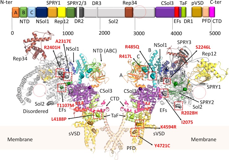
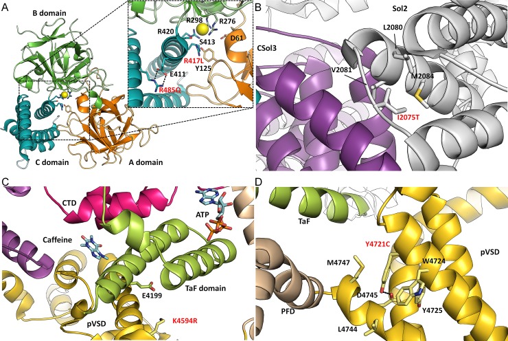
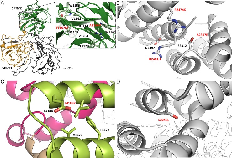
Methods: The phenotypes, genotypes and outcomes of patients in the Pediatric and Congenital Electrophysiology Society CPVT Registry with ≥2 variants in genes linked to CPVT were ascertained. The American College of Medical Genetics & Genomics (ACMG) criteria and structural mapping were used to predict the pathogenicity of variants (3D model of pig RyR2 in open-state).

Results: Among 237 CPVT subjects, 193 (81%) had genetic testing. Fifteen patients (8%) with a median age of 9 years (IQR 5-12) had ≥2 variants. Sudden cardiac arrest occurred in 11 children (73%), although none died during a median follow-up of 4.3 years (IQR 2.5-6.1). Thirteen patients (80%) had at least two RYR2 variants, while the remaining two patients had RYR2 variants plus variants in other CPVT-linked genes. Among all variants identified, re-classification of the commercial laboratory interpretation using ACMG criteria led to the upgrade from variant of unknown significance (VUS) to pathogenic/likely pathogenic (P/LP) for 5 variants, and downgrade from P/LP to VUS for 6 variants. For RYR2 variants, 3D mapping using the RyR2 model suggested that 2 VUS by ACMG criteria were P/LP, while 2 variants were downgraded to likely benign.

Conclusions: This severely affected cohort demonstrates that a minority of CPVT cases are related to ≥2 variants, which may have implications on family-based genetic counselling. While multi-variant CPVT patients were at high-risk for sudden cardiac arrest, there are insufficient data to conclude that this genetic phenomenon has prognostic implications at present. Further research is needed to determine the significance and generalizability of this observation. This study also shows that a rigorous approach to variant re-classification using the ACMG criteria and 3D mapping is important in reaching an accurate diagnosis, especially in the multi-variant population.


Conflict of interest statement

Drs. Roston & Sanatani are consultants for Audentes Therapeutics. These do not alter our adherence to PLOS ONE policies on sharing data and materials. No other authors have relevant competing interests.

Figures

Fig 1
Fig 1. Domain architecture of the cardiac RyR2.
Domains are coloured according to the linear sequence scheme shown above the ribbon diagram. Structure obtained from PDB: 5GOA. [15] The ribbon diagram shows a RyR2 dimer from the side. Red circles represent large alpha solenoid regions which are unstructured in the cryo-electron micrograph (CryoEM) structure. All variants discussed in the manuscript are shown in sphere representations. Red squares highlight location of the most likely damaging variants (expanded in Fig 2). Domains are named according to nomenclature used by des Georges, et al., 2016. A (dark orange), B (green), and C (light blue) domains: form part of the N-terminal domain (NTD); NSol (dark blue): alpha-solenoid region near the NTD; SPRY1 (light orange), SPRY2 (dark green), and SPRY3 (dark grey): three domains named after splA kinase and RyRs where they were first identified; Sol2 (light grey): second alpha-solenoid region centrally located on RyR2; Rep12 (yellow) and Rep23 (brown): four repeats (~100 aa each) in two tandem arrangements, Repeats 1 and 2 located between SPRY 1 and SPRY2, and Repeats 3 and 4 located within Sol2; CSol3 (magenta): third alpha-solenoid region located near the C-terminal; EFs (red): pair of EF hand-like motifs located within CSol3 region; TaF (light green): thumb and forefingers domain; DR1/2/3 (white): evolutionary divergent regions of RyR isoforms (not shown in the ribbon diagrams); pVSD (gold): pseudo voltage-sensing domain; PFD (wheat): pore-forming domain; CTD (pink): C-terminal domain.
Fig 2
Fig 2. Location of CPVT-associated variants on RyR2 structure.
Enlarged view of the insets are presented to highlight location of the variants and nearby residues/secondary structures. Domains are coloured according to the linear sequence scheme shown in Fig 1. All variants occur in highly conserved regions of all three RyR isoforms. Unless specified, the structures are based on the 4.2 Å resolution CryoEM open-state structure of RyR2 from porcine heart[15], PDB: 5GOA. (A) RYR2-p.R417L and RYR2-p.R485Q mutants are located centrally within RyR2 ABC domains, near the anion binding site. Residues involved in the formation of the Cl- pocket are highlighted. R485 forms a salt bridge interaction with nearby E411 residue. (B) The RYR2-p.I2075T is located within a flexible linker, joining two alpha-helices together in the Sol2 region. The Ile is surrounded by nearby hydrophobic residues. (C) The RYR2-p.K4594R variant is located in the pVSD domain, near the TaF domain. Mutation to Arg, may potentially form a salt bridge with a nearby E4199 located on the TaF domain. In RyR1 open-state structure[20] the K4594 is located near activating ligands such as caffeine and ATP, PDB: 5TAQ. (D) The RYR2-p.Y4721C variant is located within the transmembrane region of pVSD, and unsurprisingly the Y4721 is surrounded by other hydrophobic residues. Y4721 is potentially forming a polar contact with carboxyl group D4745.
Fig 3
Fig 3. Location of CPVT-associated variants on RyR2 structure (continued).
(A) RYR2-p.T1107M and p.A1136V mutations are located within the SPRY2 domain, where they are buried and surrounded by hydrophobic residues. (B) The RYR2-p.R2474K, R2401H, and RYR2-A2317E mutants are clustered within the central alpha-solenoid region of RyR2 (Sol2). The R2401 and R2474 potentially interacts with nearby D2397 and S2312 respectively. These mutations are in close proximity to the CSol3 region and the unstructured region of Sol2. This region is intrinsically flexible and generally poorly resolved in the CryoEM map. (C) RYR2-p.L4188P mutation is located in the TaF domain where it is surrounded by hydrophobic residues. (D) RYR2-p.S2246L mutation is located in the better resolved region of Sol2, where the side chain is tightly packed against neighbouring alpha helix.

References

    1. Priori SG, Wilde AA, Horie M, Cho Y, Behr ER, Berul C, et al. HRS/EHRA/APHRS expert consensus statement on the diagnosis and management of patients with inherited primary arrhythmia syndromes. Heart Rhythm. 2013;10(12):1932–63. 10.1016/j.hrthm.2013.05.014 - DOI - PubMed
    1. Swan H, Amarouch MY, Leinonen J, Marjamaa A, Kucera JP, Laitinen-Forsblom PJ, et al. Gain-of-function mutation of the SCN5A gene causes exercise-induced polymorphic ventricular arrhythmias. Circulation Cardiovascular Genetics. 2014;7(6):771–81. 10.1161/CIRCGENETICS.114.000703 - DOI - PubMed
    1. Lieve KV, Verkerk AO, Podliesna S, van der Werf C, Tanck MW, Hofman N, et al. Gain-of-function mutation in SCN5A causes ventricular arrhythmias and early onset atrial fibrillation. International Journal of Cardiology. 2017;236:187–93. 10.1016/j.ijcard.2017.01.113 - DOI - PubMed
    1. Nyegaard M, Overgaard MT, Sondergaard MT, Vranas M, Behr ER, Hildebrandt LL, et al. Mutations in calmodulin cause ventricular tachycardia and sudden cardiac death. American Journal of Human Genetics. 2012;91(4):703–12. 10.1016/j.ajhg.2012.08.015 - DOI - PMC - PubMed
    1. Gomez-Hurtado N, Boczek NJ, Kryshtal DO, Johnson CN, Sun J, Nitu FR, et al. Novel CPVT-Associated Calmodulin Mutation in CALM3 (CALM3-A103V) Activates Arrhythmogenic Ca Waves and Sparks. Circulation Arrhythmia and Electrophysiology. 2016;9(8). - PMC - PubMed

Publication types

Substances