. 2018 Nov 7;13(11):e0206143.
doi: 10.1371/journal.pone.0206143. eCollection 2018.

Primary structures of different isoforms of buffalo pregnancy-associated glycoproteins (BuPAGs) during early pregnancy and elucidation of the 3-dimensional structure of the most abundant isoform BuPAG 7

Affiliations

Primary structures of different isoforms of buffalo pregnancy-associated glycoproteins (BuPAGs) during early pregnancy and elucidation of the 3-dimensional structure of the most abundant isoform BuPAG 7

Masoud Lotfan et al. PLoS One. .

Abstract

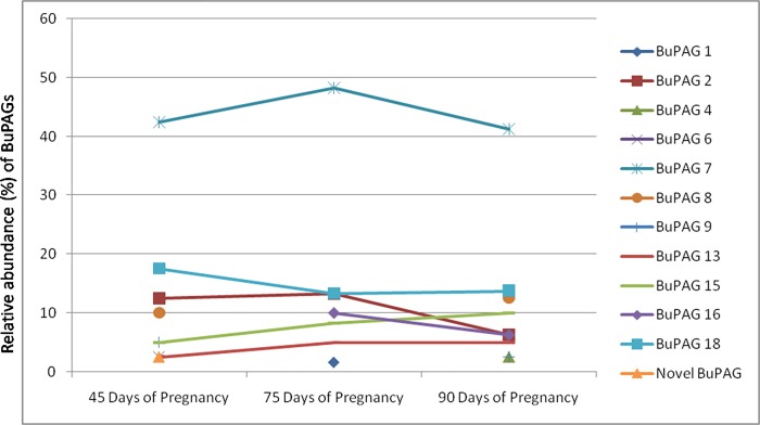
Pregnancy-associated glycoproteins (PAGs) are expressed during pregnancy by the trophoectodermal cells of fetus. Presence of PAGs in dam's circulation has been widely used in pregnancy diagnosis. The present study reports the identification and characterization of different PAG isoforms in buffalo during early stages of pregnancy. The PAG mRNAs isolated from fetal cotyledons (Pregnancy stages: 45, 75 and 90 days) were successfully cloned in pJET1.2 vector and transformed in E. coli. A total of 360 random clones were sequenced and correlated with their stages of expression. A total of 12 isoforms namely, BuPAG 1, 2, 4, 6, 7, 8, 9, 13, 15, 16, 18 and one new isoform were identified. BuPAG 7 was found as the most abundant isoform in all three stages followed by BuPAG 18. Further, a large number of variants were found for most of these isoforms. Phylogenetic relationship of identified BuPAGs showed that BuPAG 2 belonged to an ancient group while other members clustered with modern group. Three-dimensional (3D) structure of BuPAG 7 was determined by homology modeling and molecular dynamic (MD) simulations which displayed a typical fold represented by other aspartic proteinase (AP) family members. Molecular docking of Pepstatin inhibitor with BuPAG 7 revealed to interact through various hydrogen bonding and hydrophobic interactions. Various amino acid substitutions were observed in peptide-binding cleft of BuPAG 7. Superimposition of BuPAG 7 with homologous structures revealed the presence of a 35-41 amino acid long insertion (alpha helix connected by two loops) near the N- terminus which seems to be a unique feature of BuPAG 7 in AP family. This is the first report on identification and sequence characterization of PAG isoforms in buffalo with unique finding that these isoforms represent many transcript variants. We also report 3D structure of the most abundant isoform BuPAG 7 for the first time.


Conflict of interest statement

The authors have declared that no competing interests exist.

Figures

Fig 1
Fig 1. Trend analysis and relative abundance of BuPAGs.
The figure shows the trend analysis of different BuPAG isoforms and their relative abundance across three stages of early pregnancy i.e. 45 days, 75 days and 90 days.
Fig 2
Fig 2. Multiple alignment: Sequence alignment of deduced amino acid sequences of different BuPAG isoforms.
The residues in boxes differ from the consensus.
Fig 3
Fig 3. Evolutionary relationships among BuPAGs, bovine PAGs and other aspartic proteinases.
The tree was created from the deduced amino acid sequences by the Neighbor Joining method in the MEGA 4.0 program. The tree was drawn to scale, and the numbers on the branches represent the confidence levels obtained from the bootstrap analysis (1000 replicates). The tree shows the separation of BuPAGs into two groups i.e. the modern and the ancient group.
Fig 4
Fig 4
RMSD and RMSF calculations for BuPAG 7 model structure (A) Cα RMSD during 30 ns atomistic simulation run showing the structure stability and simulation integrity. (B) Observed RMSF per residue of BuPAG 7 over the 30 ns trajectory, which indicates the structural rigidity and measures the flexibility of the polypeptide chain. The region between 16±43 residues shows considerable flexibility.
Fig 5
Fig 5
(A) Ramachandran plot of the φ–ψ distribution of modeled BuPAG 7. The red, brown and yellow regions represent the most favored, additionally allowed and generously allowed regions as defined by ProCheck. (B) The overall structural display of BuPAG 7 in cartoon representation. The figure shows N- and C- terminal lobes and a central domain connecting the two lobes. The substrate binding cleft is shown between the two lobes. α- helices are shown in purple and β- strands in cyan and connecting loops in the wheat color. Three disulphides (Cys 87- Cys 92, Cys 246- Cys 250 and Cys 288- Cys 322) are shown in orange sticks. (C) The catalytic center of BuPAG 7. The conserved key catalytic residues i.e. Asp 32 and Asp 215 (pepsin numbering) are shown in orange. Other conserved residues involved in the catalytic mechanism i.e. Ser 35, Trp 39 and Tyr 75 are shown in yellow. The single amino acid substitution i.e. Ser 218 in BuPAG 7 in place of Thr 218 in pepsin and other homologs is colored in pink. The active site flap over the catalytic site is indicated by the square bracket. (D) Fireman’s Grip. The conserved residues forming the Fireman’s Grip in BuPAG 7 for structural rigidity of catalytic center. (E) Active site flap. Superimposed active site flap of BuPAG 7 (cyan) over human pepsin (Magenta) showing a slight variation in the conformation of tip region.
Fig 6
Fig 6. Substrate-binding pocket and interactions of BuPAG 7 with pepstatin.
(A) The overall structure of BuPAG 7 in complex with pepstatin in its substrate-binding pocket. Pepstatin is shown in ball and sticks representation (cyan) and interacting residues in sticks representation (green). (B) Residues involved in hydrogen bonding and other interactions with the side chains of pepstatin in sticks representation (cyan). (C) A schematic representation of interactions of pepstatin with BuPAG 7 generated using ligplot. Interactions involve both hydrogen bonding and hydrophobic contacts. Hydrogen bonds are indicated by dashed lines (green) between the atoms involved, while hydrophobic contacts are represented by an arc (red) with spokes radiating towards the ligand atoms they contact. The contacted atoms are shown with spokes radiating back.
Fig 7
Fig 7. Structural comparison with other aspartyl proteases.
(A) Superposition of pepstatin structures bound to BuPAG 7 (cyan), human pepsin (orange) and bovine chymosin (green). Pepstatin adopts an almost similar conformation in BuPAG 7 except for the extreme ends. (B) The superimposed structures of BuPAG 7, Human pepsin (1PSO), porcine pepsin (4PEP), bovine chymosin (4AUC) and human renin (3D91). The longer insertion region (35–41) in BuPAG 7 protruding out of the N-terminal lobe as compared to others is shown in circle. The change in conformation of a shorter loop is indicted by an arrow.

Similar articles

Cited by

References

    1. Zoli AP, Demez P, Beckers JF, Reznik M, Beckers A. Light and electron microscopic immunolocalization of bovine pregnancy-associated glycoprotein in the bovine placentome. Biol Reprod. 1992a;46: 623–629. - PubMed
    1. Zoli AP, Guilbault LA, Delabaut P, Orates WEB, Beckers JF. Radioimmunoassay of a bovine pregnancy-associated glycoprotein in serum: its application for pregnancy diagnosis. Biol Reprod. 1992b;46: 83–92. - PubMed
    1. Xie S, Low BG, Nagel RJ, Beckers JF, Roberts RM. A novel glycoprotein of the aspartic proteinase gene family expressed in bovine placental trophectoderm. Biol Reprod. 1994;51: 1145–1153. - PubMed
    1. Green JA, Xie S, Quan X, Bao B, Gan X, Mathialagan N, et al. Pregnancy associated glycoproteins exhibit spatially and temporally distinct expression patterns during pregnancy. Biol Reprod. 2000;62: 1624–1631. - PubMed
    1. Weems YS, Lammoglia MA, Vera-Avila HR, Randel RD, King C, Sasser RG, et al. Effect of luteinizing hormone (LH), PGE2, 8-EPI-PGE1, 8-EPI-PGE2, trichosanthin, and pregnancy specific protein B (PSPB) on secretion of progesterone in vitro by corpora lutea (CL) from nonpregnant and pregnant cows. Prostaglandins Other Lipid Mediat. 1998;55(1): 27–42. - PubMed

Publication types