. 2019 Feb 15;28(4):662-674.
doi: 10.1093/hmg/ddy383.

Identification of the novel Ido1 imprinted locus and its potential epigenetic role in pregnancy loss

Affiliations

Identification of the novel Ido1 imprinted locus and its potential epigenetic role in pregnancy loss

Philip Spinelli et al. Hum Mol Genet. .

Abstract

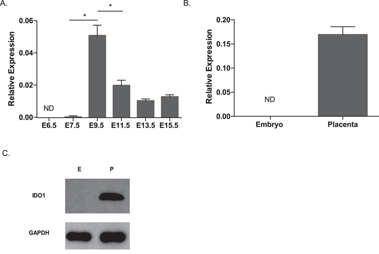
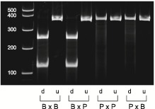
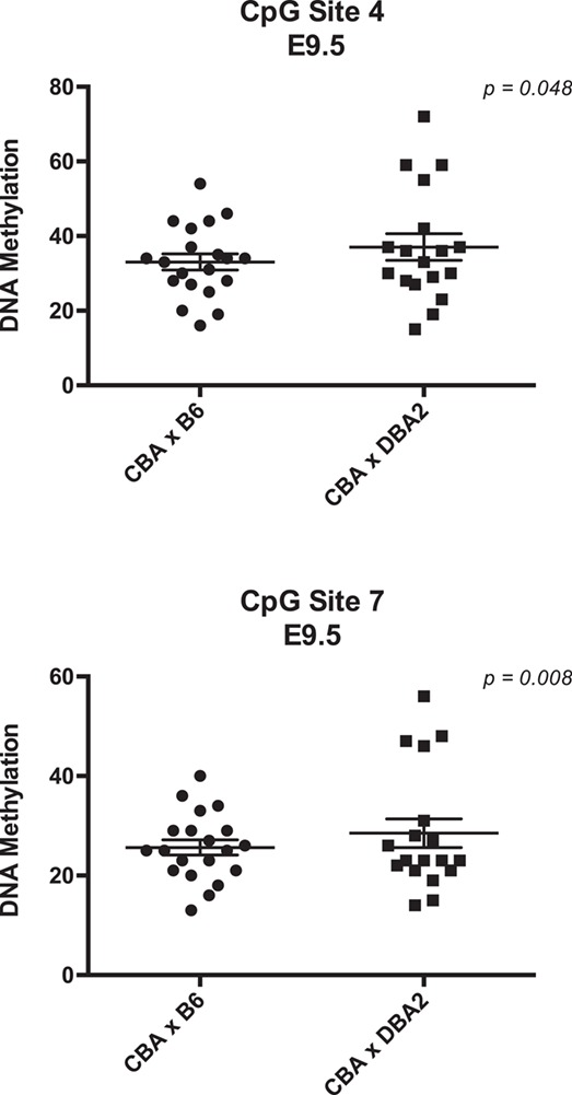
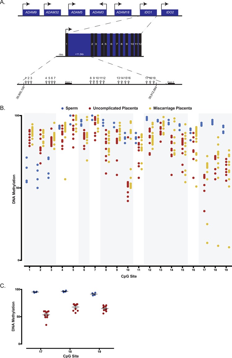
Previous studies show that aberrant tryptophan catabolism reduces maternal immune tolerance and adversely impacts pregnancy outcomes. Tryptophan depletion in pregnancy is facilitated by increased activity of tryptophan-depleting enzymes [i.e. the indolamine-2,3 dioxygenase (IDO)1 and IDO2) in the placenta. In mice, inhibition of IDO1 activity during pregnancy results in fetal loss; however, despite its important role, regulation of Ido1 gene transcription is unknown. The current study shows that the Ido1 and Ido2 genes are imprinted and maternally expressed in mouse placentas. DNA methylation analysis demonstrates that nine CpG sites at the Ido1 promoter constitute a differentially methylated region that is highly methylated in sperm but unmethylated in oocytes. Bisulfite cloning sequencing analysis shows that the paternal allele is hypermethylated while the maternal allele shows low levels of methylation in E9.5 placenta. Further study in E9.5 placentas from the CBA/J X DBA/2 spontaneous abortion mouse model reveals that aberrant methylation of Ido1 is linked to pregnancy loss. DNA methylation analysis in humans shows that IDO1 is hypermethylated in human sperm but partially methylated in placentas, suggesting similar methylation patterns to mouse. Importantly, analysis in euploid placentas from first trimester pregnancy loss reveals that IDO1 methylation significantly differs between the two placenta cohorts, with most CpG sites showing increased percent of methylation in miscarriage placentas. Our study suggests that DNA methylation is linked to regulation of Ido1/IDO1 expression and altered Ido1/IDO1 DNA methylation can adversely influence pregnancy outcomes.


Figures

Figure 1
Figure 1
Measurement of mRNA and protein expression of the Ido1 gene in mouse placenta. RT-qPCR analysis was performed to measure Ido1 mRNA expression (A) between E6.5 to E15.5, and (B) in the E9.5 embryo and placentas. Samples tested in the E6.5 and E7.5 groups were whole conceptus and ectoplacental cone, respectively. For the time course study, 4–11 conceptuses from three dams per time point were analyzed. For the tissue-specific study, 4–5 conceptuses from two dams were analyzed. Ido1 gene expression measurement was normalized to expression of the reference genes, Arpp0 and Gapdh. All values in A and B were normalized to the housekeeping genes. No significant differences were detected among E11.5–15.5 samples. (C) Western Blot analysis of E9.5 embryos and placenta using GAPDH as a loading control. Result represented analysis of three independent conceptuses. Data were analyzed using one-way ANOVA followed by Dunnett’s multiple comparison test (A) or student t-test (B) *P < 0.05 for all statistical data. Error bars represent standard error of the mean (SEM).
Figure 2
Figure 2
Restriction fragment length polymorphism analysis to show the allele-specific expression of Ido1 in E9.5 placentas. The Ido1 gene was amplified using PCR followed by restriction digest analysis. For each cross, we included both the digested (`d’) and undigested (`u’) PCR products. The B6-specific restriction fragments are 111 and 254 base pair (bp), while the PWD-specific fragment is 365 bp. Lane 1 is a 1 kb + ladder. Lanes 2–3 are B6 female mated to B6 male (B × B), lanes 4–5; B6 female mated to PWD male (B × P), lanes 6–7; P × P and lanes 8–9; P × B. Quantification of the digested samples revealed that the maternal allele contributes to 99.3 and 93.9% of total Ido1 expression in the BXP and PXB crosses, respectively. The figure is a representative of five independent experiments involving analysis of six mice per cross in each experiment.
Figure 3
Figure 3
DNA methylation analysis of the mouse Ido1 locus using bisulfite pyrosequencing and cloning sequencing. (A) Schematic of the mouse Ido1 locus on chromosome 8 showing the full-length 12.8 kb transcript containing 10 exons. Unfilled light grey lollipops represent the 37 CpG sites assayed within the mouse Ido1 locus. Arrows designate direction of gene expression. Shaded region in grey highlights the DMR. (B) Methylation levels of the 37 CpG sites in E9.5 placentas (red), sperm (blue) and oocytes (green). Each data point represents average DNA methylation from five mice. (C) Average DNA methylation levels for CpG sites 2 to 10 in E9.5 placentas (n = 5), sperm (n = 5) and oocytes (n = 5). (D) Published genome-wide ChIP-seq data set at Ido1 locus using sperm replicate 2 (19). In sperm, Ido1 is depleted of H3K4me3 but relatively enriched in H3K27me3. Shaded region represents the Ido1 DMR. (E) Ido1 DMR methylation profile at CpG sites 2–7 of E9.5 placenta and embryo, and gametes by bisulfite cloning sequencing. The gametes demonstrate differential methylation with oocytes hypomethylated (0%) and sperm hypermethylated (100%). The placenta shows similar differential between maternal (28.0%) versus paternal (72.5%) alleles, while the embryo is hypermethylated on both alleles (97.3 and 97.1%, respectively). Placenta and embryos were generated from B6xPWD and PWDxB6 hybrid crosses, while gametes are of B6 origin. Each circle represents an individual CpG site, while each row represents an independent original DNA strand. The solid and open circles represent methylated and unmethylated CpG sites, respectively. Maternal and paternal alleles were distinguished by SNPs between B6 and PWD mice. CpG site 6 was excluded from the analysis due to an existing SNP between B6 (C) and PWD (T) making it indistinguishable to determine if the CpG was unmethylated (`TG’) or a strand of PWD origin (`TG’). All error bars represent SEM. Data were analyzed using ANOVA followed by Dunnett’s multiple comparison test. P < 0.05 for all statistical analysis.
Figure 4
Figure 4
Analysis of DNA methylation of Ido1 in a spontaneous abortion mouse model. Distribution of methylation levels for CBA X DBA2 (n = 18) and CBA X B6 (n = 20) at CpG sites 4 and 7. Each data point represents one placenta. All error bars represent SEM. Data were analyzed using F variance ratio test. P < 0.05 for all statistical analysis.
Figure 5
Figure 5
DNA methylation analysis of human sperm and first trimester placentas from uncomplicated and pregnancy loss using bisulfite pyrosequencing. (A) Schematic representation of the human IDO1 locus on chromosome 8 containing 12 exons, and of the 19 CpG sites assayed within the human IDO1 locus. Arrows designate direction of gene expression. (B) Average DNA methylation levels for human sperm, uncomplicated first trimester placentas and euploid first trimester miscarriage placentas for all 19 CpG sites. A total of four sperm samples, 10 uncomplicated placentas and 13 miscarriage placentas are analyzed and presented. Global analysis of average CpG sites reveals significant differences in methylation between sperm and uncomplicated placentas. Individual CpG sites analysis shows that CpG sites 1–3, 6, 8–10, 12–15 and 17–19 significantly differed between sperm and uncomplicated placentas (regions shaded in grey). Comparing uncomplicated to miscarriage placentas, average methylation significantly differ when the CpG sites are analyzed globally. (C) Average DNA methylation of CpG sites 17, 18 and 19 in human sperm (blue) and placentas from uncomplicated pregnancies (red). Each data point represents an individual. CpG sites represent a candidate imprinted DMR as they are highly methylated in sperm but partially methylated in the placentas. Differences in percent or variance in methylation for individual CpGs were assessed using a t-test or homogeneity test of variance, and the corresponding P-values were adjusted for multiple comparisons using the Benjamini–Hochberg method. P-values and FDR of <0.05 were considered to be statistically significant.

Similar articles

Cited by

References

    1. Alegre E., López A.S., Díaz-Lagares A. and González A. (2008) Study of the plasmatic levels of tryptophan and kynurenine throughout pregnancy. Clin. Chim. Acta, 393, 132–133. - PubMed
    1. Aluvihare V.R., Kallikourdis M. and Betz A.G. (2004) Regulatory T cells mediate maternal tolerance to the fetus. Nat. Immunol., 5, 266–271. - PubMed
    1. Arefayene M., Philips S., Cao D., Mamidipalli S., Desta Z., Flockhart D.A., Wilkes D.S. and Skaar T.C. (2009) Identification of genetic variants in the human indoamine 2,3-dioxygenase (IDO1) gene, which have altered enzyme activity. Pharmacogenet. Genomics, 19 (6), 464–476. - PubMed
    1. Babak T., DeVeale B., Tsang E.K., Zhou Y., Li X., Smith K.S., Kukurba K.R., Zhang R., Li J.B., Kooy D. et al. (2015) Genetic conflict reflected in tissue-specific maps of genomic imprinting in human and mouse. Nat. Genet., 47, 544–549. - PMC - PubMed
    1. Baban B., Chandler P., McCool D., Marshall B., Munn D.H. and Mellor A.L. (2004) Indoleamine 2,3-dioxygenase expression is restricted to fetal trophoblast giant cells during murine gestation and is maternal genome specific. J. Reprod. Immunol., 61, 67–77. - PubMed

Publication types