Analysis of Inner Ear Anomalies in Unilateral Congenital Aural Atresia Combined With Microtia
- PMID: 30403837
- PMCID: PMC6453793
- DOI: 10.21053/ceo.2018.00857
Analysis of Inner Ear Anomalies in Unilateral Congenital Aural Atresia Combined With Microtia
Abstract
Objectives: The aim of this study was to analyze the incidence of inner ear anomalies in patients with unilateral congenital aural atresia (CAA) combined with microtia.
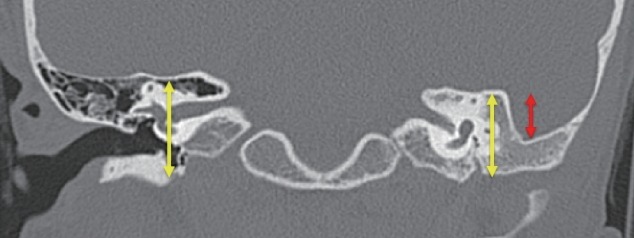
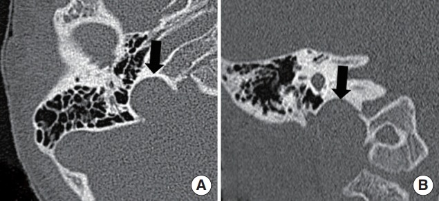
Methods: We retrospectively reviewed 61 patients with unilateral CAA combined with microtia who underwent high-resolution temporal bone computed tomography (TBCT) and hearing examination. Inner ear anomalies were analyzed using TBCT and evaluated according to the Jahrsdoerfer grading system, Marx classification, and extent of inferior displacement of the mastoid tegmen.
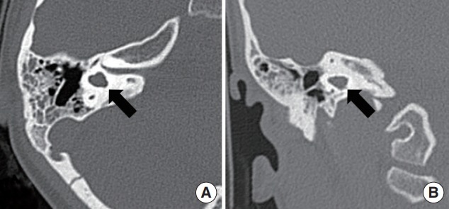
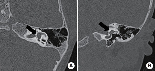
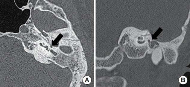
Results: Inner ear anomalies were observed in 14 patients (23.0%). Lateral semicircular canal (LSCC) dysplasia was the most common inner ear anomaly, with an incidence of 16.4%. The incidence was significantly higher on the pathologic side than on the unaffected side (P=0.002). All vascular anomalies were observed in the high-riding jugular bulb, with an incidence of 24.6%. The incidence was significantly higher on the pathologic side than on the unaffected side (P<0.001). LSCC dysplasia was significantly more common in patients with a lower Jahrsdoerfer score (odds ratio, 0.66; P=0.004).
Conclusion: The incidence of inner ear anomalies was relatively high in patients with unilateral CAA combined with microtia; LSCC dysplasia was the most common anomaly and the probability of coexistence was higher in patients with a lower Jahrsdoerfer score.
Keywords: Anomalies; Aural Atresia, Congenital; Congenital Microtia; Inner Ear; Semicircular Canals.
Conflict of interest statement
No potential conflict of interest relevant to this article was reported.
Figures
References
-
- Schuknecht HF. Congenital aural atresia. Laryngoscope. 1989 Sep;99(9):908–17. - PubMed
-
- Genc S, Kahraman E, Ozel HE, Arslan IB, Demir A, Selcuk A. Microtia and congenital aural atresia. J Craniofac Surg. 2012 Nov;23(6):1733–5. - PubMed
-
- Vrabec JT, Lin JW. Inner ear anomalies in congenital aural atresia. Otol Neurotol. 2010 Dec;31(9):1421–6. - PubMed
-
- Yuen HY, Ahuja AT, Wong KT, Yue V, van Hasselt AC. Computed tomography of common congenital lesions of the temporal bone. Clin Radiol. 2003 Sep;58(9):687–93. - PubMed