. 2018;9(1):1669-1684.
doi: 10.1080/21505594.2018.1535785.

Human monocyte-derived macrophages inhibit HCMV spread independent of classical antiviral cytokines

Affiliations

Human monocyte-derived macrophages inhibit HCMV spread independent of classical antiviral cytokines

Jennifer Becker et al. Virulence. 2018.

Abstract

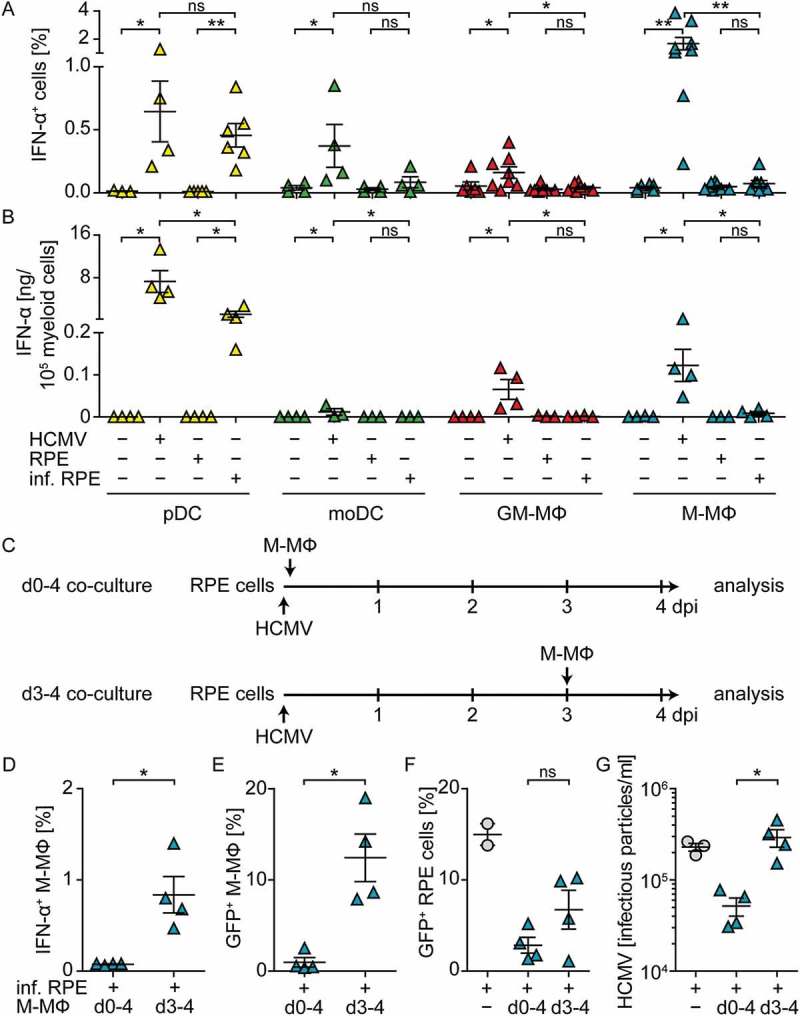
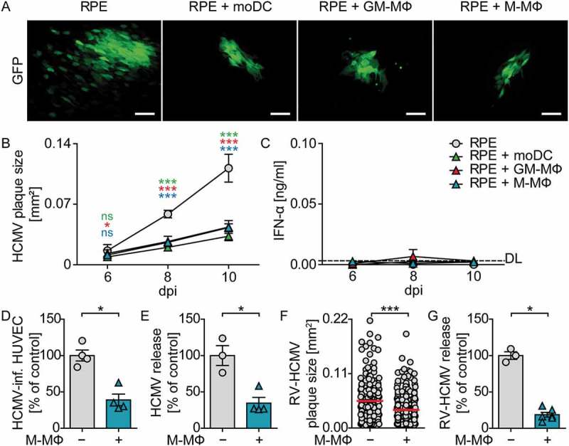
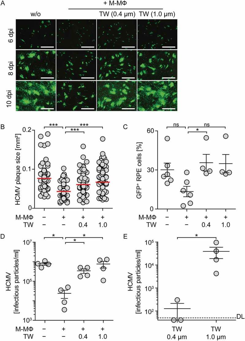
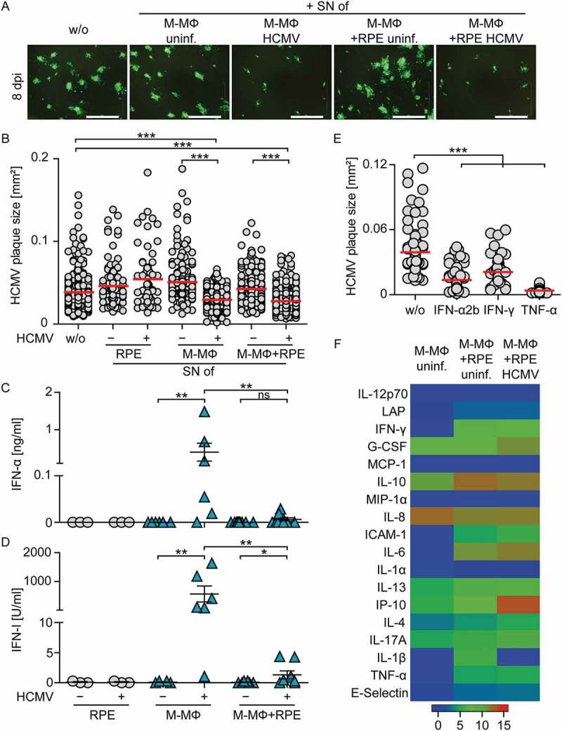
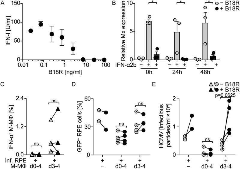
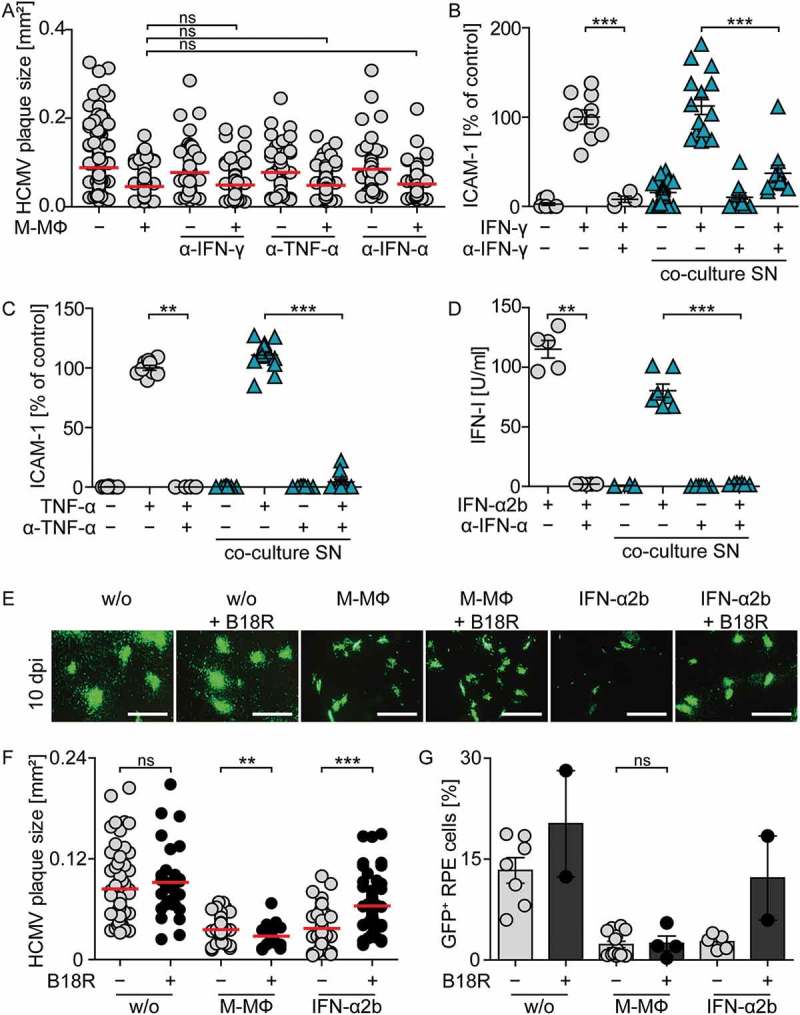
Infection of healthy individuals with human cytomegalovirus (HCMV) is usually unnoticed and results in life-long latency, whereas HCMV reactivation as well as infection of newborns or immunocompromised patients can cause life-threatening disease. To better understand HCMV pathogenesis we studied mechanisms that restrict HCMV spread. We discovered that HCMV-infected cells can directly trigger plasmacytoid dendritic cells (pDC) to mount antiviral type I interferon (IFN-I) responses, even in the absence of cell-free virus. In contrast, monocyte-derived cells only expressed IFN-I when stimulated by cell-free HCMV, or upon encounter of HCMV-infected cells that already produced cell-free virus. Nevertheless, also in the absence of cell-free virus, i.e., upon co-culture of infected epithelial/endothelial cells and monocyte-derived macrophages (moMΦ) or dendritic cells (moDC), antiviral responses were induced that limited HCMV spread. The induction of this antiviral effect was dependent on cell-cell contact, whereas cell-free supernatants from co-culture experiments also inhibited virus spread, implying that soluble factors were critically needed. Interestingly, the antiviral effect was independent of IFN-γ, TNF-α, and IFN-I as indicated by cytokine inhibition experiments using neutralizing antibodies or the vaccinia virus-derived soluble IFN-I binding protein B18R, which traps human IFN-α and IFN-β. In conclusion, our results indicate that human macrophages and dendritic cells can limit HCMV spread by IFN-I dependent as well as independent mechanisms, whereas the latter ones might be particularly relevant for the restriction of HCMV transmission via cell-to-cell spread.

Keywords: Human cytomegalovirus; epithelial cells; macrophages; plasmacytoid dendritic cells; type I interferons.


Figures

Figure 1.
Figure 1.
Co-culture of myeloid cells with HCMV-infected RPE cells induces enhanced IFN-α responses by pDC, but not by monocyte-derived cells. pDC, moDC, GM-CSF MΦ (GM-MΦ), or M-CSF MΦ (M-MΦ) were directly infected with HCMV-GFP at MOI 3. Furthermore, RPE cells were infected with HCMV at MOI 12, washed to remove cell-free virus, and co-cultured with myeloid cell subsets at a 1:4 ratio for 24 h and (a) percentages of intracellular IFN-α+ myeloid cells or (b) the IFN-α content in cell-free supernatants was determined by flow cytometry or an ELISA method, respectively. (c) RPE cells were infected with HCMV-GFP (MOI 12), washed, and co-cultured with M-CSF MΦ either from day (d) 0–4 post infection or from d 3–4 post infection and (d) percentages of intracellular IFN-α+ M-CSF MΦ, (e) HCMV-GFP+ M-CSF MΦ, (f) HCMV-GFP+ RPE cells, or (g) infectious HCMV particles in the supernatant of such cultures were determined. Mean ± SEM of (a) 4–8, (b) 3–4, or (d-g) 4 different donors from 2–4 independent experiments. inf. = infected, ns = not significant, *: p ≤ 0.05, **: p ≤ 0.01 (one-tailed Mann-Whitney test).
Figure 2.
Figure 2.
Co-culture of monocyte-derived cells with HCMV-infected RPE cells reduces viral spread. RPE cells were infected with HCMV-GFP at MOI 0.1, washed, and co-cultured with moDC, GM-CSF MΦ, or M-CSF MΦ at a 1:4 ratio. (a) 10 dpi HCMV plaque formation was analyzed by fluorescence microscopy (scale bar = 100 µm). (b) 6, 8, and 10 dpi the size of individual HCMV plaques was determined by using ImageJ software. (c) Secreted IFN-α was monitored by ELISA analysis of cell-free supernatants. HUVEC were infected with HCMV-GFP at MOI 1, co-cultured with M-CSF MΦ, and 10 dpi (d) HUVEC were analyzed for percentages of GFP expressing cells by flow cytometry and (e) supernatants were analyzed for the amount of infectious virus particles. RPE cells were infected with RV-TB40-BACKL7-SE-EGFP (RV-HCMV), co-cultured with M-CSF MΦ, and 13 dpi (f) the size of individual HCMV plaques was determined and (g) supernatants were analyzed for the amount of infectious virus particles. Data in (d/e/g) show values as percent of control infections in HUVEC or RPE mono-cultures. Mean ±SEM of (b/c) 4–7 ((b) 26–102 plaques analyzed), (d/e) 4, and (f/g) 6 ((f) 212–368 plaques analyzed) different donors from 2–3 independent experiments. DL = detection limit, dpi = days post infection, *: p ≤ 0.05, ***: p ≤ 0.001 (one-tailed Mann-Whitney test).
Figure 3.
Figure 3.
Inhibition of direct cell-cell contact between macrophages and infected epithelial cells impairs protection against HCMV. RPE cells were infected with HCMV-GFP at MOI 0.1, washed, and co-cultured with M-CSF MΦ at a 1:2 ratio. M-CSF MΦ were added directly onto RPE cells or into transwell inserts with pore sizes of 0.4 µm or 1.0 µm. (a) 6, 8, and 10 dpi HCMV plaque formation was analyzed by fluorescence microscopy (scale bar = 1 mm) and 10 dpi (b) the size of individual HCMV plaques or (c) percentage of HCMV-GFP+ RPE cells was determined. Numbers of infectious HCMV particles were determined 10 dpi in (d) co-cultures or in the RPE cell compartment of the transwell co-cultures as well as (e) in the transwell inserts, which contained the M-CSF MΦ. Mean size of (b) 77–119 plaques using 4 different donors or mean ±SEM of (c-e) 3–4 different donors from 2 independent experiments. DL = detection limit, ns = not significant, *: p ≤ 0.05, ***: p ≤ 0.001 (one-tailed Mann-Whitney).
Figure 4.
Figure 4.
Supernatants from co-cultures of M-CSF MΦ and HCMV-infected RPE cells inhibit viral spread. Cell-free supernatants from uninfected or HCMV-infected RPE (MOI 12) or M-CSF-MΦ (MOI 3) mono-cultures and RPE/M-CSF-MΦ co-cultures were harvested 2 dpi and transferred onto fresh RPE cells infected with HCMV-GFP at MOI 0.1. (a) 8 dpi virus plaque formation was analyzed by fluorescence microscopy (scale bar = 1 mm) and (b) the size of individual plaques was determined. Cell-free supernatants prepared as described above were analyzed for (c) secreted IFN-α and (d) IFN-I activity using an ELISA method or an Mx2-Luc reporter cell line, respectively. (e) RPE cells were infected with HCMV-GFP at MOI 0.1 and incubated with recombinant (rec.) IFN-α2b, IFN-γ, or TNF-α for 8 d. Then the plaque size was analyzed. (f) Cell-free supernatants prepared as described above were analyzed for different secreted proteins using a bead-based cytokine array. Data visualize log (2) values of the measured cytokine concentrations in [pg/ml]. Mean size of (b) 67–371 plaques using 6 different donors or (e) 49–100 plaques from 3 independent experiments. Mean ±SEM of (c/d) 6 different donors from 3 independent experiments or data of (f) 3 different donors from 2 independent experiments. SN = supernatant, uninf. = uninfected, *: p ≤ 0.05, **: p ≤ 0.01, ***: p ≤ 0.001 (one-tailed Mann-Whitney test).
Figure 5.
Figure 5.
Co-cultures of macrophages and infected RPE cells incubated for four days show IFN-I independent inhibition of HCMV propagation. (a) Different concentrations of B18R were added to rec. IFN-α2b [100 U/ml] and after 1 h incubation the IFN-I activity was determined using an Mx2-Luc reporter cell line. (b) Co-cultures of M-CSF-MΦ and HCMV-infected RPE cells were set up, treated with 100 ng/ml B18R, and 0, 24, and 48 h later cultures were stimulated with 100 U/ml rec. IFN-α2b for 4 h and then Mx induction was analyzed by qPCR. (c-e) RPE cells were infected with HCMV-GFP at MOI 12, washed, and co-cultured with M-CSF MΦ from d 0–4 or d 3–4 post infection with (black symbols) or without (grey symbols) the daily addition of 100 ng/ml B18R. (c) Percentages of intracellular IFN-α+ M-CSF-MΦ, (d) HCMV-GFP+ RPE cells, or (e) infectious HCMV particles in the supernatants of such cultures were determined. Mean ±SEM of (a) 3 independent experiments and (b) 3 or (c/d/e) 4 different donors from 2 independent experiments. inf. = infected, *: p ≤ 0.05 (one-tailed Mann-Whitney test, (b)), ns = not significant (p ≥ 0.29) (one-tailed Wilcoxon signed rank test (c/d/e)).
Figure 6.
Figure 6.
Inhibition of IFN-γ, TNF-α, or IFN-I does not impair protection against HCMV in co-cultures of macrophages and infected RPE cells. RPE cells were infected with HCMV-GFP at MOI 0.1, washed, and co-cultured with M-CSF MΦ with or without the addition of antibodies against human IFN-γ, TNF-α, or the IFN-I receptor. 10 dpi co-culture supernatants were harvested and (a) HCMV plaque formation was analyzed by fluorescence microscopy. To determine the remaining neutralizing potential of anti-human IFN-γ or TNF-α antibodies in day 10 co-culture supernatants, RPE cells were treated with (b) 100 U/ml rec. IFN-γ or (c) 10 ng/ml rec. TNF-α in the presence or absence of anti-human IFN-γ or TNF-α antibodies or the harvested day 10 co-culture supernatants, respectively, and upregulation of ICAM-1 expression was analyzed by flow cytometry. Data are shown as percent of ICAM-1 upregulation after IFN-γ or TNF-α treatment of RPE cells (control). (d) To determine the remaining potential of anti-human IFNAR antibodies to block IFN-I responses in day 10 co-culture supernatants, Mx2-Luc reporter cells were treated with 100 U/ml rec. IFN-α2b in the presence or absence of anti-human IFNAR antibodies or day 10 co-culture supernatants and IFN-I activity was measured. RPE cells were infected with HCMV-GFP at MOI 0.1, washed, and co-cultured with M-CSF MΦ with or without the addition of 100 ng/ml B18R daily. Furthermore, infected RPE cells were treated with 100 U/ml rec. IFN-α2b which was pretreated for 1 h with 100 ng/ml B18R. 10 dpi (e) HCMV plaque formation was analyzed by fluorescence microscopy (scale bar = 1 mm) and (f) the size of individual HCMV plaques or (g) percentages of HCMV-GFP+ RPE cells were determined. Mean size of (a) 116–184 or (f) 32–68 plaques using 2–7 different donors or mean ± SEM of (b/c) 7, (d) 5, and (g) 4 different donors from 2–3 independent experiments. ns = not significant, **: p ≤ 0.01, ***: p ≤ 0.001 (one-tailed Mann-Whitney test).

Similar articles

Cited by

References

    1. Griffiths P, Baraniak I, Reeves M.. The pathogenesis of human cytomegalovirus. J Pathol. 2015;235:288–297. - PubMed
    1. Revello MG, Gerna G. Diagnosis and management of human cytomegalovirus infection in the mother, fetus, and newborn infant. Clin Microbiol Rev. 2002;15:680–715. - PMC - PubMed
    1. Sinclair J, Sissons P. Latency and reactivation of human cytomegalovirus. J Gen Virol. 2006;87:1763–1779. - PubMed
    1. Pawelec G. Immunosenenescence: role of cytomegalovirus. Exp Gerontol. 2014;54:1–5. - PubMed
    1. Loewendorf A, Benedict CA. Modulation of host innate and adaptive immune defenses by cytomegalovirus: timing is everything. J Intern Med. 2010;267:483–501. - PMC - PubMed

Publication types