

Affinity-Bead-Mediated Enrichment of CD8+ Lymphocytes from Peripheral Blood Progenitor Cell Products Using Acoustophoresis
- PMID: 30404275
- PMCID: PMC6190086
- DOI: 10.3390/mi7060101
Affinity-Bead-Mediated Enrichment of CD8+ Lymphocytes from Peripheral Blood Progenitor Cell Products Using Acoustophoresis
Abstract
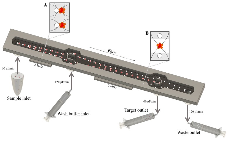
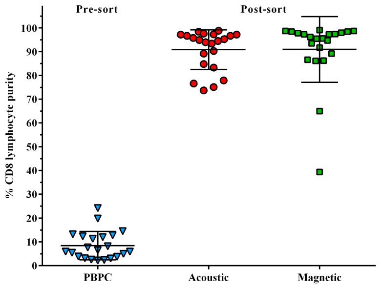
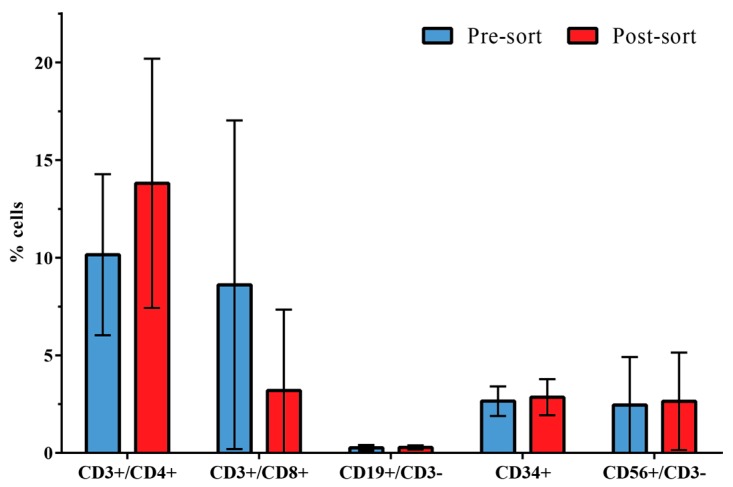
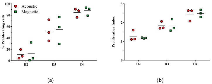
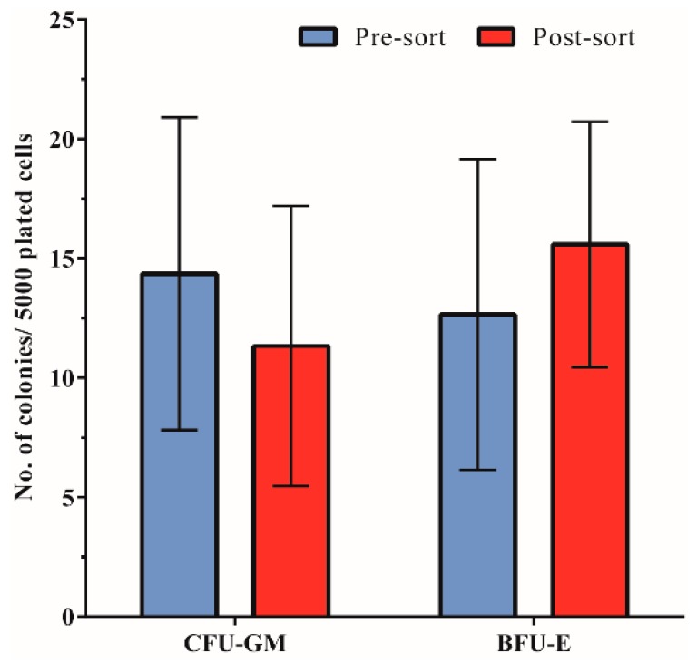
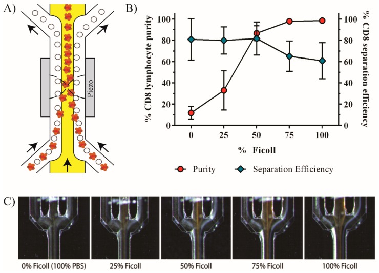
Acoustophoresis is a technique that applies ultrasonic standing wave forces in a microchannel to sort cells depending on their physical properties in relation to the surrounding media. Cell handling and separation for research and clinical applications aims to efficiently separate specific cell populations. Here, we investigated the sorting of CD8 lymphocytes from peripheral blood progenitor cell (PBPC) products by affinity-bead-mediated acoustophoresis. PBPC samples were obtained from healthy donors (n = 4) and patients (n = 18). Mononuclear cells were labeled with anti-CD8-coated magnetic beads and sorted on an acoustophoretic microfluidic device and by standard magnetic cell sorting as a reference method. CD8 lymphocytes were acoustically sorted with a mean purity of 91% ± 8% and a median separation efficiency of 63% (range 15.1%⁻90.5%) as compared to magnetic sorting (purity 91% ± 14%, recovery 29% (range 5.1%⁻47.3%)). The viability as well as the proliferation capacity of sorted lymphocytes in the target fraction were unimpaired and, furthermore, hematopoietic progenitor cell assay revealed a preserved clonogenic capacity post-sorting. Bead-mediated acoustophoresis can, therefore, be utilized to efficiently sort less frequent CD8+ lymphocytes from PBPC products in a continuous flow mode while maintaining cell viability and functional capacity of both target and non-target fractions.
Keywords: CD8 lymphocytes; PBPC; acoustophoresis; cell sorting; magnetic-beads; peripheral blood progenitor cells; ultrasound.
Conflict of interest statement
Thomas Laurell and Stefan Scheding are board members, founders and shareholders of AcouSort AB which develops acoustophoresis technology. The work presented in the current paper is not biased by commercial interest from AcouSort AB.
Figures
Similar articles
-
Efficient purification of CD4+ lymphocytes from peripheral blood progenitor cell products using affinity bead acoustophoresis.Cytometry A. 2014 Nov;85(11):933-41. doi: 10.1002/cyto.a.22507. Epub 2014 Jul 22. Cytometry A. 2014. PMID: 25053536
-
Efficient removal of platelets from peripheral blood progenitor cell products using a novel micro-chip based acoustophoretic platform.PLoS One. 2011;6(8):e23074. doi: 10.1371/journal.pone.0023074. Epub 2011 Aug 9. PLoS One. 2011. PMID: 21857996 Free PMC article.
-
Label-free neuroblastoma cell separation from hematopoietic progenitor cell products using acoustophoresis - towards cell processing of complex biological samples.Sci Rep. 2019 Jun 19;9(1):8777. doi: 10.1038/s41598-019-45182-3. Sci Rep. 2019. PMID: 31217534 Free PMC article.
-
Label-free separation of neuroblastoma patient-derived xenograft (PDX) cells from hematopoietic progenitor cell products by acoustophoresis.Stem Cell Res Ther. 2021 Oct 15;12(1):542. doi: 10.1186/s13287-021-02612-2. Stem Cell Res Ther. 2021. PMID: 34654486 Free PMC article.
-
[Collection of hematopoietic progenitor cells from healthy donors].Acta Med Croatica. 2009 Jun;63(3):237-44. Acta Med Croatica. 2009. PMID: 19827352 Review. Croatian.
Cited by
-
Acoustic enrichment of heterogenous circulating tumor cells and clusters from patients with metastatic prostate cancer.medRxiv [Preprint]. 2023 Dec 4:2023.12.04.23299128. doi: 10.1101/2023.12.04.23299128. medRxiv. 2023. Update in: Anal Chem. 2024 May 7;96(18):6914-6921. doi: 10.1021/acs.analchem.3c05371. PMID: 38106097 Free PMC article. Updated. Preprint.
-
Acoustic Enrichment of Heterogeneous Circulating Tumor Cells and Clusters from Metastatic Prostate Cancer Patients.Anal Chem. 2024 May 7;96(18):6914-6921. doi: 10.1021/acs.analchem.3c05371. Epub 2024 Apr 24. Anal Chem. 2024. PMID: 38655666 Free PMC article.
-
Aptamer Affinity-Bead Mediated Capture and Displacement of Gram-Negative Bacteria Using Acoustophoresis.Micromachines (Basel). 2019 Nov 11;10(11):770. doi: 10.3390/mi10110770. Micromachines (Basel). 2019. PMID: 31718045 Free PMC article.
-
Three-Dimensional Reservoir-Based Dielectrophoresis (rDEP) for Enhanced Particle Enrichment.Micromachines (Basel). 2018 Mar 10;9(3):123. doi: 10.3390/mi9030123. Micromachines (Basel). 2018. PMID: 30424057 Free PMC article.
-
Potential of the acoustic micromanipulation technologies for biomedical research.Biomicrofluidics. 2021 Nov 24;15(6):061301. doi: 10.1063/5.0073596. eCollection 2021 Dec. Biomicrofluidics. 2021. PMID: 34849184 Free PMC article.
References
-
- Gratwohl A., Baldomero H., Horisberger B., Schmid C., Passweg J., Urbano-Ispizua A. Accreditation Committee of the European Group for Blood and Marrow Transplantation (EBMT) Current trends in hematopoietic stem cell transplantation in Europe. Blood. 2002;100:2374–2386. doi: 10.1182/blood-2002-03-0675. - DOI - PubMed
-
- Dreger P., Haferlach T., Eckstein V., Jacobs S., Suttorp M., Löffler H., Müller-Ruchholtz W., Schmitz N. G-CSF-mobilized peripheral blood progenitor cells for allogeneic transplantation: Safety, kinetics of mobilization, and composition of the graft. Br. J. Haematol. 1994;87:609–613. doi: 10.1111/j.1365-2141.1994.tb08321.x. - DOI - PubMed
-
- Theilgaard-Mönch K., Raaschou-Jensen K., Palm H., Schjødt K., Heilmann C., Vindeløv L., Jacobsen N., Dickmeiss E. Flow cytometric assessment of lymphocyte subsets, lymphoid progenitors, and hematopoietic stem cells in allogeneic stem cell grafts. Bone Marrow Transplant. 2001;28:1073–1082. doi: 10.1038/sj.bmt.1703270. - DOI - PubMed
Grants and funding
LinkOut - more resources
Full Text Sources
Other Literature Sources
Research Materials
