Review
. 2016 Oct 2;7(10):179.
doi: 10.3390/mi7100179.

Neural Probes for Chronic Applications

Affiliations
Review

Neural Probes for Chronic Applications

Geon Kook et al. Micromachines (Basel). .

Abstract

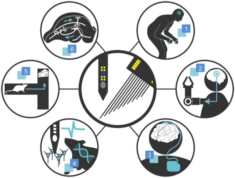
Developed over approximately half a century, neural probe technology is now a mature technology in terms of its fabrication technology and serves as a practical alternative to the traditional microwires for extracellular recording. Through extensive exploration of fabrication methods, structural shapes, materials, and stimulation functionalities, neural probes are now denser, more functional and reliable. Thus, applications of neural probes are not limited to extracellular recording, brain-machine interface, and deep brain stimulation, but also include a wide range of new applications such as brain mapping, restoration of neuronal functions, and investigation of brain disorders. However, the biggest limitation of the current neural probe technology is chronic reliability; neural probes that record with high fidelity in acute settings often fail to function reliably in chronic settings. While chronic viability is imperative for both clinical uses and animal experiments, achieving one is a major technological challenge due to the chronic foreign body response to the implant. Thus, this review aims to outline the factors that potentially affect chronic recording in chronological order of implantation, summarize the methods proposed to minimize each factor, and provide a performance comparison of the neural probes developed for chronic applications.

Keywords: biocompatibility; biocompatible coating; chronic implant; foreign body response; neural probe; neural recording.


Conflict of interest statement

The authors declare no conflict of interest.

Figures

Figure 1
Figure 1
Graphical overview of applications of the neural probe technology. 1. Parkinson’s Diseases; 2. Neuroprosthetics; 3. Brain Pacemaker; 4. Investigation of Brain Diseases; 5. Cognitive Experiments; 6. Brain Mapping.
Figure 2
Figure 2
Optical photographs of the state-of-the-art neural probes: (a) high density (top: 3D array from Normann et al. [24]; bottom: 200-electrode shank from Scholvin et al. [17]); (b) functionalities (top: micro-LED for optogenetics from Wu et al. [13]; bottom: mixer-integrated microfluidic channel for drug delivery from Shin et al. [11]); (c) integrated circuits (top: a 64-channel integrated circuit (IC) with wireless transmission from Sodagra et al. [19]; bottom: a 52-channel IC from Lopez et al. [18]), and (d-e) biocompatibility ((d): syringe-injectable flexible 3D probe from Liu et al. [62]; (e): dissolvable silk-coated probe from Wu et al. [35]). Standard neural probes consist of single or multiple shanks and microelectrode arrays integrated at the end of the shanks for neural recording; The syringe-injectable probe shown in (d) is a new type of neural probe which provides 3D access to the brain with minimal damage. All figures reprinted with permission.
Figure 3
Figure 3
(a) Graphical representations of common surface modification strategies, reprinted with permission from Marin et al. [81]. An optimal surface should consist of an insulation layer that facilitates the adsorption of proteins, adhesion of fibroblast, and adhesion of neurons and glial cells without macrophage reaction and a microelectrode surface that attracts neurons without adhesion of fibroblasts or macrophage reaction; (b) Graphical representation of selective coatings of different bioactive materials on a neural probe, reprinted with permission from Abidian et al. [116]. Drug-loaded biodegradable nanofibers are encapsulated by a biocompatible alginate hydrogel as the insulation layer and poly(3,4-ethylenedioxythiophen) (PEDOT) is electrochemically polymerized on the microelectrode.
Figure 4
Figure 4
3D macroporous brain probes reprinted with permission from Xie et al. Schematics of (a) insertion scheme and (b) 3D spherical structure with flexible arms that are designed to protrude through the glial scar and form a close contact to the neurons; and (c) immunohistochemistry (IHC) of the insertion site demonstrating an extremely low immune response with a high neuronal density [85].

References

    1. Calabresi P., Centonze D., Bernardi G. Electrophysiology of dopamine in normal and denervated striatal neurons. Trends Neurosci. 2000;23:S57–S63. doi: 10.1016/S1471-1931(00)00017-3. - DOI - PubMed
    1. Altuna A., Bellistri E., Cid E., Aivar P., Gal B., Berganzo J., Gabriel G., Guimerà A., Villa R., Fernández L.J. SU-8 based microprobes for simultaneous neural depth recording and drug delivery in the brain. Lab Chip. 2013;13:1422–1430. doi: 10.1039/c3lc41364k. - DOI - PubMed
    1. Alivisatos A.P., Chun M., Church G.M., Greenspan R.J., Roukes M.L., Yuste R. The brain activity map project and the challenge of functional connectomics. Neuron. 2012;74:970–974. doi: 10.1016/j.neuron.2012.06.006. - DOI - PMC - PubMed
    1. Scanziani M., Häusser M. Electrophysiology in the age of light. Nature. 2009;461:930–939. doi: 10.1038/nature08540. - DOI - PubMed
    1. Hochberg L.R., Bacher D., Jarosiewicz B., Masse N.Y., Simeral J.D., Vogel J., Haddadin S., Liu J., Cash S.S., van der Smagt P. Reach and grasp by people with tetraplegia using a neurally controlled robotic arm. Nature. 2012;485:372–375. doi: 10.1038/nature11076. - DOI - PMC - PubMed

LinkOut - more resources