. 2019;20(5):608-616.
doi: 10.1080/15384047.2018.1539289. Epub 2018 Nov 7.

Activation of CXCL5-CXCR2 axis promotes proliferation and accelerates G1 to S phase transition of papillary thyroid carcinoma cells and activates JNK and p38 pathways

Affiliations

Activation of CXCL5-CXCR2 axis promotes proliferation and accelerates G1 to S phase transition of papillary thyroid carcinoma cells and activates JNK and p38 pathways

Dong Cui et al. Cancer Biol Ther. 2019.

Abstract

C-X-C motif chemokine ligand 5 (CXCL5) is initially identified to recruit neutrophils by interacting with its receptor, C-X-C motif chemokine receptor 2 (CXCR2). Our prior work demonstrated that the expression levels of CXCL5 and CXCR2 were higher in the papillary thyroid carcinoma (PTC) tumors than that in the non-tumors. This study was performed to further investigate how this axis regulates the growth of PTC cells. B-CPAP cells (BRAFV600E) and TPC-1 cells (RET/PTC rearrangement) expressing CXCR-2 were used as in vitro cell models. Our results showed that the recombinant human CXCL5 (rhCXCL5) promoted the proliferation of PTC cells. rhCXCL5 accelerated the G1/S transition, upregulated the expression of a group of S (DNA synthesis) or M (mitosis)-promoting cyclins and cyclin-dependent kinases (CDKs), and downregulated CDK inhibitors in PTC cells. The CDS region of homo sapiens CXCL5 gene was inserted into an eukaryotic expression vector to mediate the overexpression of CXCL5 in PTC cells. The phosphorylation of c-Jun N-terminal kinases (JNK) and p38, and the nuclear translocation of c-Jun were enhanced by CXCL5 overexpression, whereas attenuated by CXCR2 antagonist SB225002. Additionally, CXCL5/CXCR2 axis, JNK and p38 pathway inhibitors, SB225002, SP600125 and SB203580, suppressed the growth of PTC cells overexpressing CXCL5 in nude mice, respectively. Collectively, our study demonstrates a growth-promoting effect of CXCL5-CXCR2 axis in PTC cells in vitro and in vivo.

Keywords: CXCL5; CXCR2; JNK pathway; cell cycle; cell proliferation; p38 pathway; papillary thyroid carcinoma.


Figures

Figure 1.
Figure 1.
Exogenous rhCXCL5 promotes PTC cell proliferation (a) The basal expression of CXCR2 in B-CPAP, TPC-1 and KTC-1 cells were determined with Western blot analysis. (b-c) B-CPAP cells and (d-e) TPC-1 cells were treated with 1, 5, 10 nM rhCXCL5 protein for 24, 48, 72 or 96 hrs before CCK-8 (n = 5), immunofluorescence and Western blot assays using anti-PCNA antibody (bars, 50 μm; 48 hrs post rhCXCL5 treatment). **P < 0.01 and *P < 0.05 (1 nM rhCXCL5 versus Control), ##P < 0.01 and #P < 0.05 (5 nM rhCXCL5 versus Control), §§P < 0.01 (10 nM rhCXCL5 versus Control). PTC, papillary thyroid carcinoma; rhCXCL5, recombinant human C-X-C motif chemokine ligand 5; CXCR2, C-X-C motif chemokine receptor 2; PCNA, proliferating cell nuclear antigen.
Figure 2.
Figure 2.
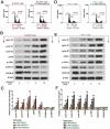
Exogenous rhCXCL5 promotes the G1 to S transition in PTC cells (a) B-CPAP cells and (D) TPC-1 cells were treated with or without 10 nM rhCXCL5 protein for 48 hrs before flow cytometry assay (Propidium staining). The levels of a group of cell cycle-associated proteins in (b-c) B-CPAP cells and (e-f) TPC-1 cells were detected with western blot (with β-actin as control; n = 3). **P < 0.01 (1 nM rhCXCL5 versus Control), ##P < 0.01 (5 nM rhCXCL5 versus Control), §§P < 0.01 (10 nM rhCXCL5 versus Control). PTC, papillary thyroid carcinoma; rhCXCL5, recombinant human C-X-C motif chemokine ligand 5; CXCR2, C-X-C motif chemokine receptor 2.
Figure 3.
Figure 3.
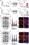
Forced overexpression of CXCL5-induced cell proliferation and activation of JNK and p38 pathways are inhibited by CXCR2 inhibitor SB225002. The complete CDS region of homo sapiens CXCL5 (NM_002994) was amplified and inserted into the eukaryotic expression vector pEGFP-N1 (CXCL5 OE plasmid). B-CPAP cells and TPC-1 cells were transfected with CXCL5 OE plasmid or negative control (NC) plasmid by using Lipofectamine™ 3000 transfection reagent, and 48 hrs later, the mRNA and protein expression levels of CXCL5 were respectively determined with (a & d) real-time quantitative PCR and (b & e) western blot. Additionally, forty-eight hours post the transfection, CXCR2 antagonist SB225002 (5 μM) was used to treat PTC cells for another 24 hrs. (c & f) Cell viability was determined with CCK-8 as well (n = 3). (g-h & j-k) The levels of nuclear c-Jun, phosphorylated or total JNK1/2, and phosphorylated or total p38 were determined with western blot analysis. (i & l) Immunofluorescence assay using anti-c-Jun antibody was performed to probe the expression of c-Jun in PTC cells (bars, 50 μm). All data were expressed in mean ± standard deviation (SD) (n = 3). **P < 0.01; *P < 0.05. CXCL5, C-X-C motif chemokine ligand 5; CXCR2, C-X-C motif chemokine receptor 2; JNK, c-Jun N-terminal kinases
Figure 4.
Figure 4.
Blockade of JNK or p38 pathway suppresses CXCL5 overexpression-induced cell proliferation. Forty-eight hours post the transfection, 10 μM SP600125 (JNK pathway inhibitor) or 20 μM SB203580 (p38 pathway inhibitor) was used to treat PTC cells for another 24 hrs, and the expression levels of PCNA, cyclin E1 and B1 were determined with western blot assay. **P < 0.01; *P < 0.05. CXCL5, C-X-C motif chemokine ligand 5; JNK, c-Jun N-terminal kinases; PCNA, proliferating cell nuclear antigen
Figure 5.
Figure 5.
Forced overexpression of CXCL5 promotes the growth of B-CPAP xenografted tumors via activation of JNK or p38 pathway. BCPAP cells were resuspended in 0.1 mL serum-free RPMI medium, mixed with 0.1 mL Metrigel and then injected into the axillary skin of nude mice. (a) The xenografted tumors were isolated and photographed on day 28 post the injection of cancer cells (left column). Immunofluorescence assay using PCNA antibody was conducted to probe PCNA-positive cells within the tumors (bars, 50 μm; middle and right columns). (b) The levels of nuclear c-Jun and phosphorylated or total p38 were determined with western blot analysis, and (c) calculated by comparing to H3 histone or β-actin. (d-f) Nude mice injected with CXCL5 OE BCPAP cells were given CXCR2 antagonist SB225002 (intraperitoneal injection of 10 mg/kg for three times a week), JNK pathway inhibitor SP600125 (intraperitoneal injection of 5 mg/kg for three times a week) or p38 inhibitor SB203580 (subcutaneous injection of 4 mg/kg for five times a week) for four weeks. The xenografted tumors were isolated and photographed on day 28 post the injection of cancer cells. The tumor volume was measured every three day before sacrificing on day 28. All data were expressed in mean ± standard error (n = 6). **P < 0.01; *P < 0.05. CXCL5, C-X-C motif chemokine ligand 5; PCNA, anti-proliferating cell nuclear antigen; OE, overexpression

References

    1. Kitahara CM, Sosa JA.. The changing incidence of thyroid cancer. Nat Rev Endocrinol. 2016;12:646–653. doi:10.1038/nrendo.2016.110. - DOI - PMC - PubMed
    1. Xing M, Haugen BR, Schlumberger M.. Progress in molecular-based management of differentiated thyroid cancer. Lancet. 2013;381:1058–1069. doi:10.1016/S0140-6736(13)60109-9. - DOI - PMC - PubMed
    1. Chang MS, McNinch J, Basu R, Simonet S. Cloning and characterization of the human neutrophil-activating peptide (ENA-78) gene. J Biol Chem. 1994;269:25277–25282. - PubMed
    1. Vandercappellen J, Van Damme J, Struyf S. The role of CXC chemokines and their receptors in cancer. Cancer Lett. 2008;267:226–244. doi:10.1016/j.canlet.2008.04.050. - DOI - PubMed
    1. Cui D, Zhao Y, Xu J. Activated CXCL5-CXCR2 axis promotes the migration, invasion and EMT of papillary thyroid carcinoma cells via modulation of beta-catenin pathway. Biochimie. 2018. doi:10.1016/j.biochi.2018.02.009. - DOI - PubMed

MeSH terms