. 2018 Nov 7;18(1):178.
doi: 10.1186/s12866-018-1310-9.

Evolution and functional characterization of pectate lyase PEL12, a member of a highly expanded Clonostachys rosea polysaccharide lyase 1 family

Affiliations

Evolution and functional characterization of pectate lyase PEL12, a member of a highly expanded Clonostachys rosea polysaccharide lyase 1 family

Lea Atanasova et al. BMC Microbiol. .

Abstract

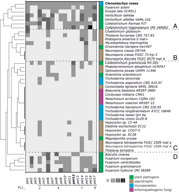
Background: Pectin is one of the major and most complex plant cell wall components that needs to be overcome by microorganisms as part of their strategies for plant invasion or nutrition. Microbial pectinolytic enzymes therefore play a significant role for plant-associated microorganisms and for the decomposition and recycling of plant organic matter. Recently, comparative studies revealed significant gene copy number expansion of the polysaccharide lyase 1 (PL1) pectin/pectate lyase gene family in the Clonostachys rosea genome, while only low numbers were found in Trichoderma species. Both of these fungal genera are widely known for their ability to parasitize and kill other fungi (mycoparasitism) and certain species are thus used for biocontrol of plant pathogenic fungi.

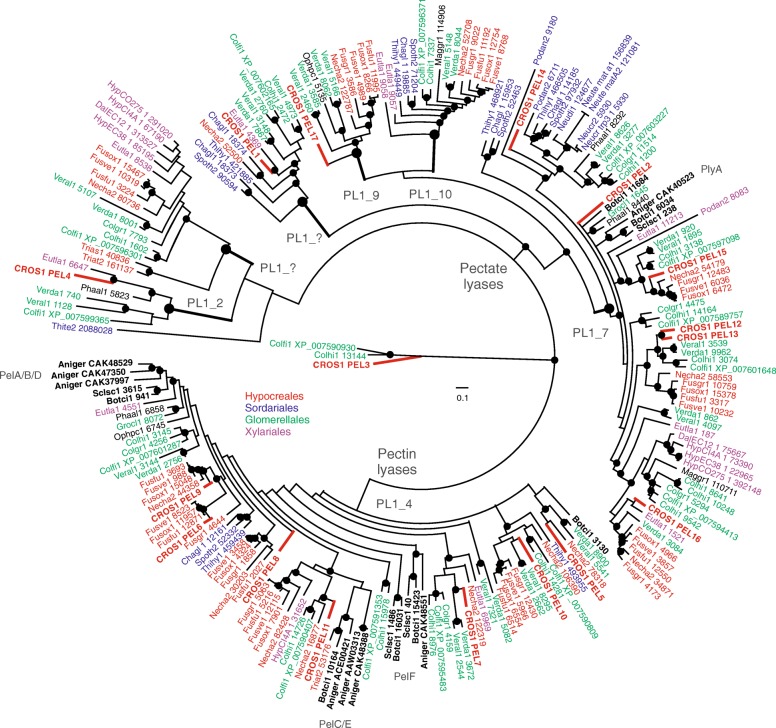
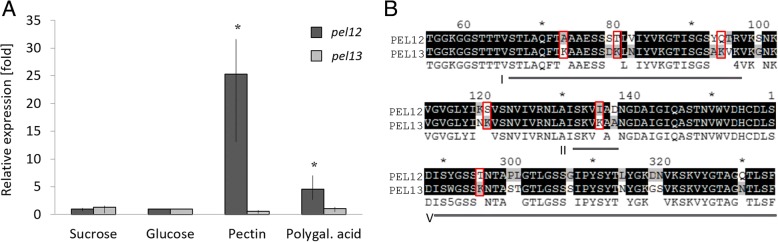
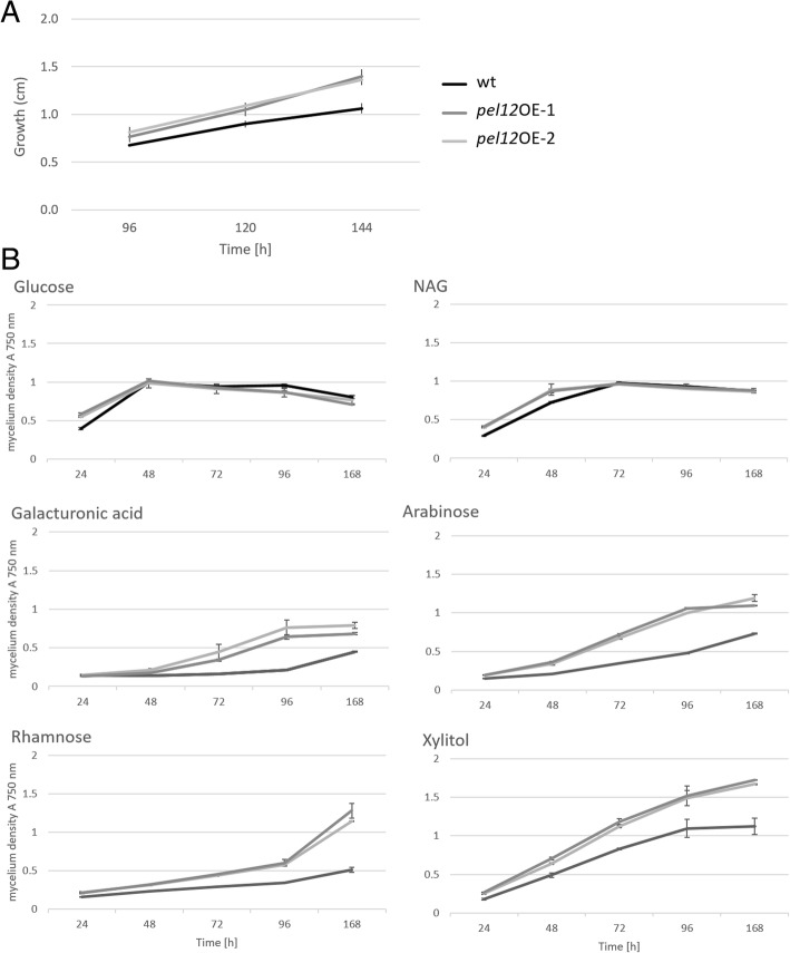
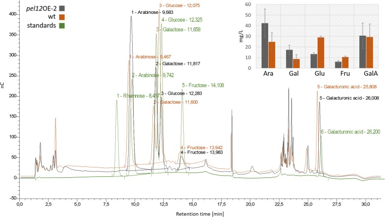
Results: In order to understand the role of the high number of pectin degrading enzymes in Clonostachys, we studied diversity and evolution of the PL1 gene family in C. rosea compared with other Sordariomycetes with varying nutritional life styles. Out of 17 members of C. rosea PL1, we could only detect two to be secreted at acidic pH. One of them, the pectate lyase pel12 gene was found to be strongly induced by pectin and, to a lower degree, by polygalacturonic acid. Heterologous expression of the PEL12 in a PL1-free background of T. reesei revealed direct enzymatic involvement of this protein in utilization of pectin at pH 5 without a requirement for Ca2+. The mutants showed increased utilization of pectin compounds, but did not increase biocontrol ability in detached leaf assay against the plant pathogen Botrytis cinerea compared to the wild type.

Conclusions: In this study, we aimed to gain insight into diversity and evolution of the PL1 gene family in C. rosea and other Sordariomycete species in relation to their nutritional modes. We show that C. rosea PL1 expansion does not correlate with its mycoparasitic nutritional mode and resembles those of strong plant pathogenic fungi. We further investigated regulation, specificity and function of the C. rosea PEL12 and show that this enzyme is directly involved in degradation of pectin and pectin-related compounds, but not in C. rosea biocontrol.

Keywords: Clonostachys rosea; Enzyme; Pectin; Phylogeny; Plant biomass degradation; Trichoderma reesei.


Conflict of interest statement

Ethics approval and consent to participate

Not applicable

Consent for publication

Not applicable

Competing interests

The authors declare that they have no competing interests.

Publisher’s Note

Springer Nature remains neutral with regard to jurisdictional claims in published maps and institutional affiliations.

Figures

Fig. 1
Fig. 1
Hierarchical clustering based on PL1 gene content in Sordariomycetes and Botrytis cinerea and Sclerotinia sclerotiorum (Leotiomycetes). The clusters are marked with letters A to D. The squares colored with four different shades of gray represent the number of C. rosea orthologs per species. The color annotation on the right marks the nutritional mode of fungi
Fig. 2
Fig. 2
Phylogenetic relationships of PL1 proteins among Sordariomycetes, A. niger (Eurotiomycetes) and S. sclerotiorum and B. cinerea (Leotiomycetes). Predicted amino acid sequences were aligned by MUSCLE and were used to construct a Bayesian phylogenetic tree implemented in MrBayes. The marked nodes represent posterior probabilities greater or equal than 95%. The species abbreviations followed by genome protein IDs are given on the tree, the full species names are listed in the Additional file 1: Table S1. Included Leotiomyces and Eurotiomycetes are marked in bold black
Fig. 3
Fig. 3
Reverse conservation analysis of PL1_7 orthologs revealed areas of putative functional divergence. a Amino acid conservation was estimated using Rate4Site, based on a MUSCLE alignment of fungal PL1_7 orthologs, and plotted as W mean scores in arbitrary units. The black and grey lines represent the closest orthologs in the PEL12/PEL13 and PEL2 clades (Additional file 2: Figure S1), respectively. b Cartoon representation of the PEL12 model. Defined regions are those that are mostly variable. I = pos. 67–98 (in red), II = pos. 131–137 (in blue), III = pos. 176–182 (in magenta), IV = pos. 217–223 (in cyan), V = pos. 287–327 (in yellow). The pentagalacturonate ligand is shown in orange sticks, and the two calcium ions positioned at two putative Ca2+ binding site, are shown in pink. In subfigure b. the catalytic base, Arg 225, is shown in green sticks. The hydrolytic cleavage of the polygalacturonic substrate will occur at the position denoted by the arrow. The pentagalacturonate and calcium ligands are extracted from a superposition of PDB:ID 3KRG
Fig. 4
Fig. 4
a Differential gene expression of the pel12 (BN869_T0006915) and pel13 (BN869_T00007653) genes in C. rosea, induced by 0.5% sucrose, glucose, pectin or polygalacturonic acid. Asterisks indicate significantly (P ≤ 0.05) higher expression compared with the glucose control treatment. b Mutational shifts from non-charged amino acids to positively charged lysine (red squares) in the PEL12 and PEL13 protein sequences. Roman numbers mark the evolutionary variable regions I, II and V detected between the closest paralogs (see Fig. 3). Asterisks mark next ten amino acids
Fig. 5
Fig. 5
Utilization of carbon sources in T. reesei QM 9414 and pel12OE mutants. a Growth of strains on 1% agarose supplemented with 2% pectin. b Growth rates of strains on selected carbon sources assessed by BIOLOG Phenotypic assay for filamentous fungi. The cultures were incubated at 28 °C in darkness
Fig. 6
Fig. 6
HPAEC-PAD analysis of T. reesei QM 9414 and the pel12OE-2 mutant supernatants collected after 72 h post inoculation on minimal medium supplemented with 1% apple pectin, pH 5. The bars represent the detected quantity of monosaccharides arabinose (Ara), galactose (Gal), glucose (Glu), fructose (Fru), and galacturonic acid (GalA) in the culture extracts. nC is the unit of electric charge measured in nano-Coulomb, meaning the charge transported by a constant current of one Ampere in one second

References

    1. Willats WG, McCartney L, Mackie W, Knox JP. Pectin: cell biology and prospects for functional analysis. Plant Mol Biol. 2001;47:9–27. doi: 10.1023/A:1010662911148. - DOI - PubMed
    1. Mohnen D. Pectin structure and biosynthesis. Curr Opin Plant Biol. 2008;11:266–277. doi: 10.1016/j.pbi.2008.03.006. - DOI - PubMed
    1. O’Neill MA, Ishii T, Albersheim P, Darvill AG. Rhamnogalacturonan II: structure and function of a borate cross-linked cell wall pectic polysaccharide. Annu Rev Plant Biol. 2004;55:109–139. doi: 10.1146/annurev.arplant.55.031903.141750. - DOI - PubMed
    1. Buchanan BB, Gruissem W, Jones RL, editors. Biochemistry & Molecular Biology of Plants. Rockville: American Society of Plant Physiologists; 2000.
    1. Müller-Maatsch J, Bencivenni M, Caligiani A, Tedeschi T, Bruggeman G, Bosch M, et al. Pectin content and composition from different food waste streams. Food Chem. 2016;201:37–45. doi: 10.1016/j.foodchem.2016.01.012. - DOI - PubMed

Publication types

MeSH terms

LinkOut - more resources