

Incidence and predictors of left ventricular thrombus by cardiovascular magnetic resonance in acute ST-segment elevation myocardial infarction treated by primary percutaneous coronary intervention: a meta-analysis
- PMID: 30404623
- PMCID: PMC6222991
- DOI: 10.1186/s12968-018-0494-3
Incidence and predictors of left ventricular thrombus by cardiovascular magnetic resonance in acute ST-segment elevation myocardial infarction treated by primary percutaneous coronary intervention: a meta-analysis
Abstract
Introduction: The incidence of left ventricular (LV) thrombus formation in ST-segment elevation myocardial infarction (STEMI) patients in the current era of primary percutaneous coronary intervention (PCI) is not well established. We performed a meta-analysis to assess the actual incidence and predictors of LV thrombus by cardiovascular magnetic resonance (CMR) in STEMI treated by primary PCI.
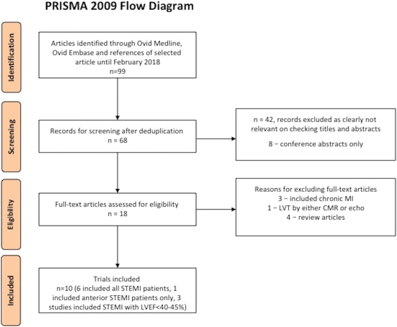
Methods: We searched MEDLINE and EMBASE databases up to February 2018. We included all studies published as a full-text article, reporting the incidence of LV thrombus by CMR within 1 month following acute STEMI in patients treated by primary PCI. A binary random-effects model was used to estimate the pooled incidence of LV thrombus. The diagnostic performance of transthoracic echocardiography (TTE) as compared with CMR was pooled to obtain the sensitivity and specificity of TTE with CMR as the gold standard. Embolic and bleeding complications of LV thrombus were also evaluated.
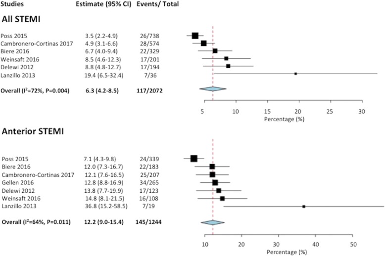
Results: Ten studies were included in the meta-analysis. The incidence of LV thrombus by CMR in all-comer STEMI patients (n = 2072) was 6.3% with 96% of LV thrombus occurring in those with anterior STEMI (12.2% incidence). When only anterior STEMI with LVEF< 50% were considered (n = 447), the incidence of LV thrombus was 19.2%. Compared with CMR, the sensitivity of TTE to detect LV thrombus was 29% with a specificity of 98%. The sensitivity of TTE increased to 70% in those with anterior STEMI and reduced LVEF. LV thrombus resolved in 88% of cases by 3 to 6 months. After 1-2 years follow-up, the embolic complication rate was similar at 1.5% (P = 0.25) but the bleeding complication rate was significantly higher (8.8% versus 0.5%, P < 0.001) in the LV thrombus group on triple therapy when compared to the no LV thrombus group on dual antiplatelet therapy.
Conclusion: In the primary PCI era, CMR detection of an LV thrombus post-STEMI remains high with incidence of nearly 20% in anterior STEMI with depressed LVEF. Patients with LV thrombus treated by triple therapy had similar embolic complications but higher bleeding complications than those with no LV thrombus treated with dual antiplatelet therapy. A 3 month follow-up CMR scan to guide anticoagulation duration might help mitigate bleeding risk.
Keywords: Cardiovascular magnetic resonance; Left ventricular thrombus; Primary percutaneous coronary intervention; ST-segment elevation myocardial infarction.
Conflict of interest statement
Ethics approval and consent to participate
Not applicable.
Consent for publication
Not applicable.
Competing interests
The authors declare that they have no competing interests.
Publisher’s Note
Springer Nature remains neutral with regard to jurisdictional claims in published maps and institutional affiliations.
Figures
References
-
- O'Gara PT, Kushner FG, Ascheim DD, et al. 2013 ACCF/AHA guideline for the management of ST-elevation myocardial infarction: a report of the American College of Cardiology Foundation/American Heart Association task force on practice guidelines. Circulation. 2013;127(4):e362–e425. - PubMed
-
- Ibanez B, James S, Agewall S, et al. 2017 ESC guidelines for the management of acute myocardial infarction in patients presenting with ST-segment elevation: the task force for the management of acute myocardial infarction in patients presenting with ST-segment elevation of the European Society of Cardiology (ESC) Eur Heart J. 2018;39(2):119–177. doi: 10.1093/eurheartj/ehx393. - DOI - PubMed
Publication types
MeSH terms
Substances
Grants and funding
LinkOut - more resources
Full Text Sources
Medical
Miscellaneous
