

Porcine epidemic diarrhea virus S1 protein is the critical inducer of apoptosis
- PMID: 30404647
- PMCID: PMC6222994
- DOI: 10.1186/s12985-018-1078-4
Porcine epidemic diarrhea virus S1 protein is the critical inducer of apoptosis
Abstract
Background: Porcine Epidemic Diarrhea (PED) is an acute and highly contagious enteric disease caused by PED virus (PEDV), characterized by vomitting, watery diarrhea and fatal dehydration with high mortality in sucking piglets of one week of age. Although PEDV induced cell apoptosis has been established in vitro and in vivo, the functional protein that contributes to this event remains unclear.
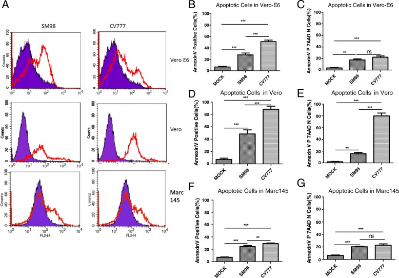
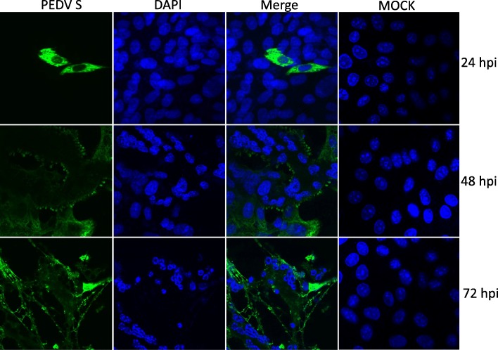
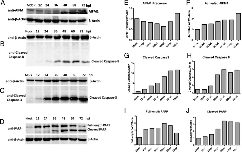
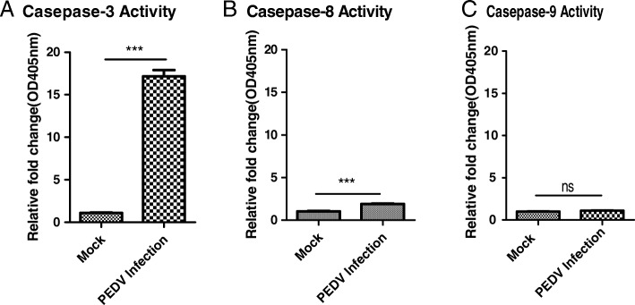
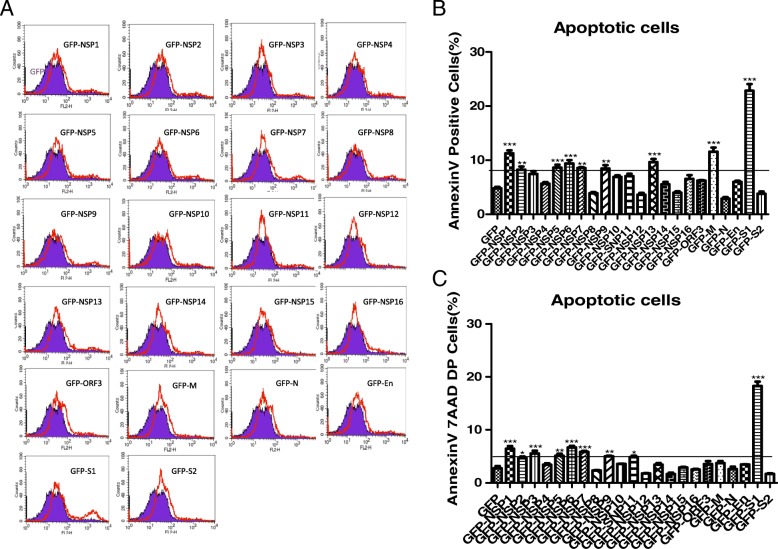
Methods: The activation or cleavage of main apoptosis-associated molecular such as AIFM1, caspase-3, caspase-8, caspase-9 and PARP in PEDV infected host cells were analyzed by western blotting. The nuclear change of infected cell was monitored by confocal immunofluorescence assay. The overexpressing plasmids of 16 non-structural proteins (Nsp1-16) and 6 structural proteins (M, N, E, ORF3, S1 and S2) were constructed by cloning. Cell apoptosis induced by PEDV or overexpression non-structural or structural proteins was measured by the flow cytometry assay.
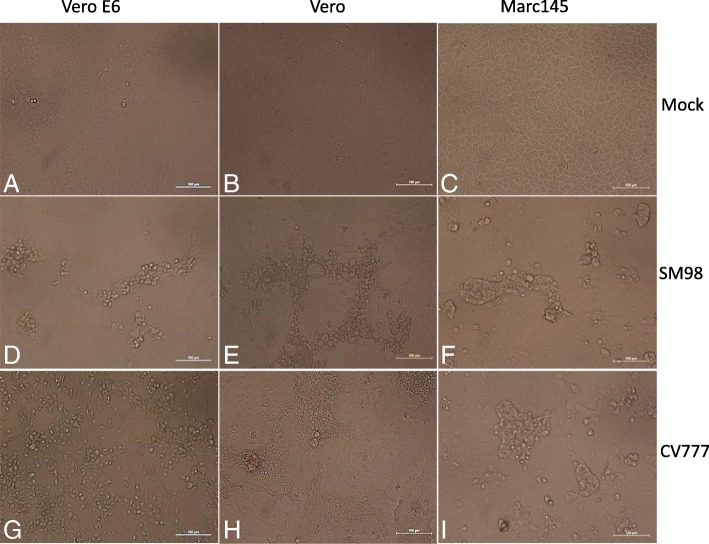
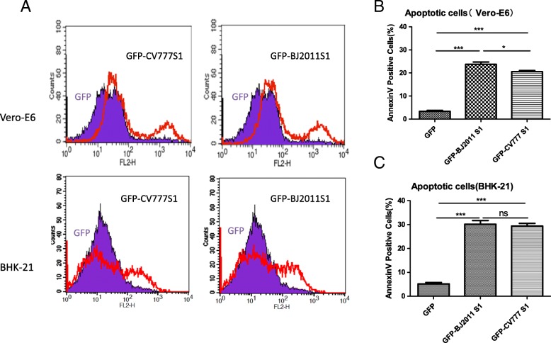
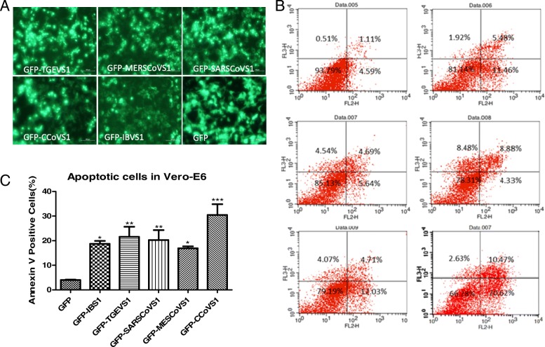
Results: PEDV could infect various host cells including Vero, Vero-E6 and Marc-145 and cause obvious cytopathic effects, including roundup, cell fusion, cell membrane vacuolation, syncytium formation and cause apparent apoptosis. In infected cells, PEDV-induced apoptosis is accompanied by nuclear concentration and fragmentation as a result of caspase-3 and caspase-8 activation and AIFM1 and PARP cleavage. Overexpression of S1 Spike protein of PEDV SM98 strain effectively induced host cell apoptosis, while the expression of the other non-structure proteins (Nsp1-16) and structural proteins (M, N, E, S2 and ORF3) has no or less effect on cell apoptosis. Similarly, expression of S1 protein from wild-type strain BJ2011 or cell-adapted strain CV777, also induce apoptosis in transfected cells. Finally, we demonstrated that the S1 proteins from various coronavirus family members such as TGEV, IBV, CCoV, SARS and MERS could also induce Vero-E6 cells apoptosis.
Conclusion: S1 Spike protein is one of the most critical functional proteins that contribute to cell apoptosis. Expression of S1 proteins of the coronavirus tested in this study could all induce cell apoptosis suggesting S1 maybe is an effective inducer in Coronavirus-induced cell apoptosis and targeting S1 protein expression probably is a promising strategy to inhibit coronavirus infection and thus mediated apoptosis on host cells.
Keywords: Apoptosis; Apoptosis-inducing factor mitochondria associated 1 (AIFM1); Porcine epidemic diarrhea virus (PEDV); Spike S1 protein.
Conflict of interest statement
Ethics approval and consent to participate
Not applicable
Consent for publication
Not applicable.
Competing interests
The authors declare that they have no conflict of interest
Publisher’s Note
Springer Nature remains neutral with regard to jurisdictional claims in published maps and institutional affiliations.
Figures
Similar articles
-
The Accessory Protein ORF3 Contributes to Porcine Epidemic Diarrhea Virus Replication by Direct Binding to the Spike Protein.Viruses. 2018 Jul 28;10(8):399. doi: 10.3390/v10080399. Viruses. 2018. PMID: 30060558 Free PMC article.
-
Deletion of a 197-Amino-Acid Region in the N-Terminal Domain of Spike Protein Attenuates Porcine Epidemic Diarrhea Virus in Piglets.J Virol. 2017 Jun 26;91(14):e00227-17. doi: 10.1128/JVI.00227-17. Print 2017 Jul 15. J Virol. 2017. PMID: 28490591 Free PMC article.
-
Characterization and evolution of the coronavirus porcine epidemic diarrhoea virus HLJBY isolated in China.Transbound Emerg Dis. 2020 Jan;67(1):65-79. doi: 10.1111/tbed.13321. Epub 2019 Aug 22. Transbound Emerg Dis. 2020. PMID: 31381232 Free PMC article.
-
Cellular entry of the porcine epidemic diarrhea virus.Virus Res. 2016 Dec 2;226:117-127. doi: 10.1016/j.virusres.2016.05.031. Epub 2016 Jun 15. Virus Res. 2016. PMID: 27317167 Free PMC article. Review.
-
[Advances in reverse genetics to treat porcine epidemic diarrhea virus].Sheng Wu Gong Cheng Xue Bao. 2017 Feb 25;33(2):205-216. doi: 10.13345/j.cjb.160282. Sheng Wu Gong Cheng Xue Bao. 2017. PMID: 28956377 Review. Chinese.
Cited by
-
Swine Enteric Coronavirus: Diverse Pathogen-Host Interactions.Int J Mol Sci. 2022 Apr 2;23(7):3953. doi: 10.3390/ijms23073953. Int J Mol Sci. 2022. PMID: 35409315 Free PMC article. Review.
-
DNAJA3 Interacts with PEDV S1 Protein and Inhibits Virus Replication by Affecting Virus Adsorption to Host Cells.Viruses. 2022 Oct 31;14(11):2413. doi: 10.3390/v14112413. Viruses. 2022. PMID: 36366511 Free PMC article.
-
Alterations of Suckling Piglet Jejunal Microbiota Due to Infection With Porcine Epidemic Diarrhea Virus and Protection Against Infection by Lactobacillus salivarius.Front Vet Sci. 2021 Dec 9;8:771411. doi: 10.3389/fvets.2021.771411. eCollection 2021. Front Vet Sci. 2021. PMID: 34957282 Free PMC article.
-
Porcine Epidemic Diarrhea Virus (PEDV) ORF3 Enhances Viral Proliferation by Inhibiting Apoptosis of Infected Cells.Viruses. 2020 Feb 14;12(2):214. doi: 10.3390/v12020214. Viruses. 2020. PMID: 32075094 Free PMC article.
-
A Comprehensive View on the Protein Functions of Porcine Epidemic Diarrhea Virus.Genes (Basel). 2024 Jan 26;15(2):165. doi: 10.3390/genes15020165. Genes (Basel). 2024. PMID: 38397155 Free PMC article. Review.
References
-
- Kusanagi K, Kuwahara H, Katoh T, Nunoya T, Ishikawa Y, Samejima T, et al. Isolation and serial propagation of porcine epidemic diarrhea virus in cell cultures and partial characterization of the isolate. The Journal of veterinary medical science / the Japanese Society of Veterinary Science. 1992;54(2):313–318. doi: 10.1292/jvms.54.313. - DOI - PubMed
-
- Lee HM, Lee BJ, Tae JH, Kweon CH, Lee YS, Park JH. Detection of porcine epidemic diarrhea virus by immunohistochemistry with recombinant antibody produced in phages. The Journal of veterinary medical science / the Japanese Society of Veterinary Science. 2000;62(3):333–337. doi: 10.1292/jvms.62.333. - DOI - PubMed
Publication types
MeSH terms
Substances
Grants and funding
LinkOut - more resources
Full Text Sources
Research Materials
Miscellaneous
