. 2019 Jan 17;93(3):e01887-18.
doi: 10.1128/JVI.01887-18. Print 2019 Feb 1.

Infection and Functional Modulation of Human Monocytes and Macrophages by Varicella-Zoster Virus

Affiliations

Infection and Functional Modulation of Human Monocytes and Macrophages by Varicella-Zoster Virus

Jarrod J Kennedy et al. J Virol. .

Abstract

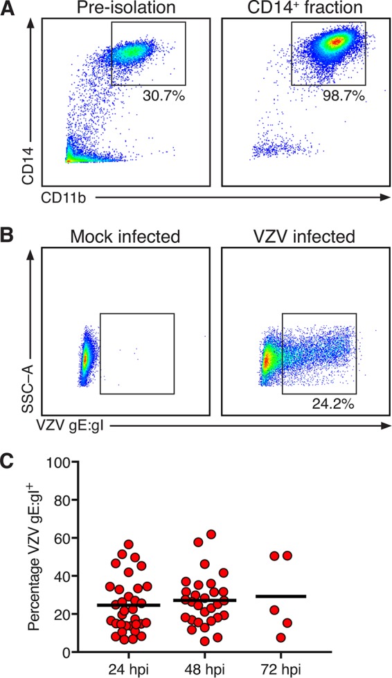
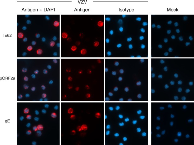
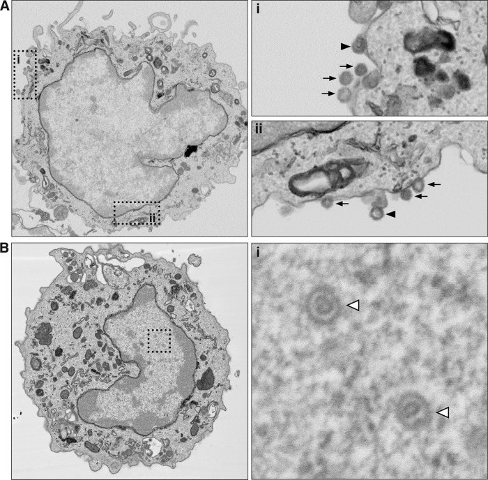
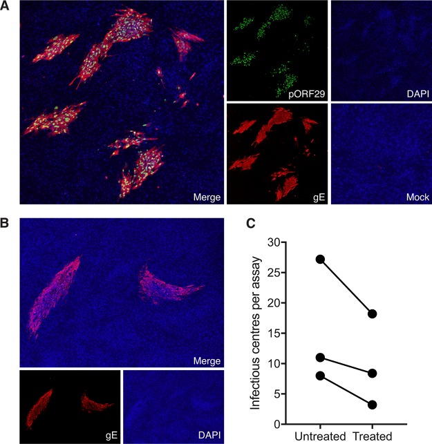
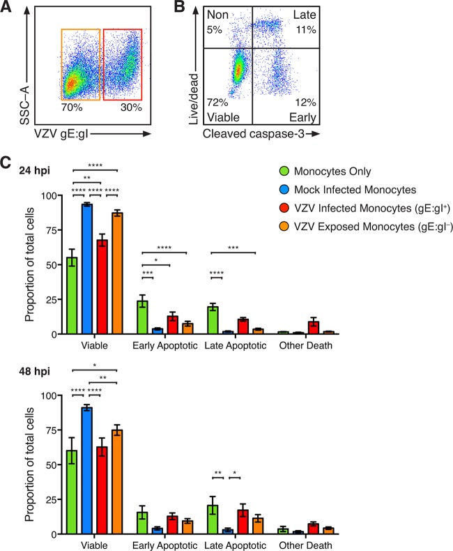
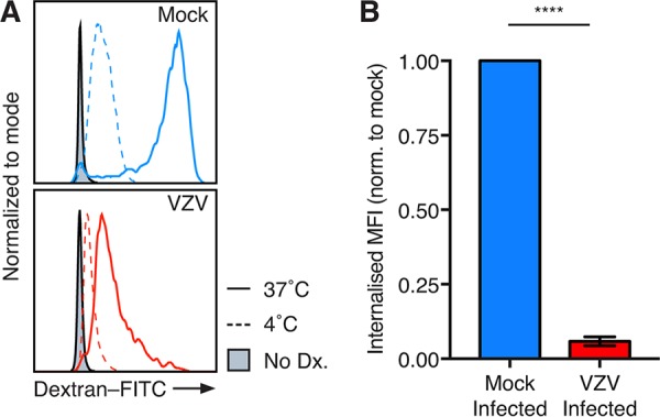
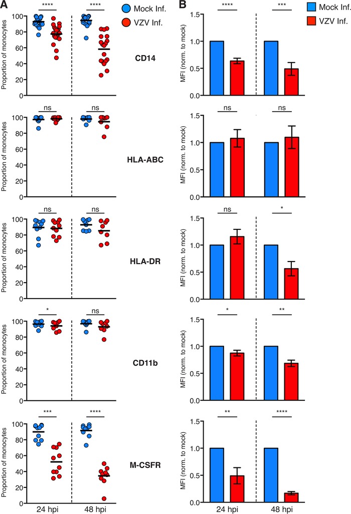
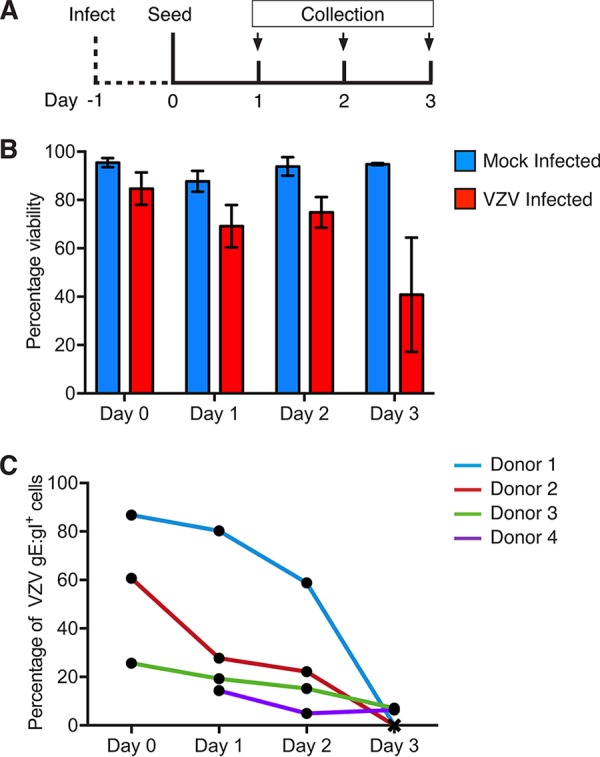
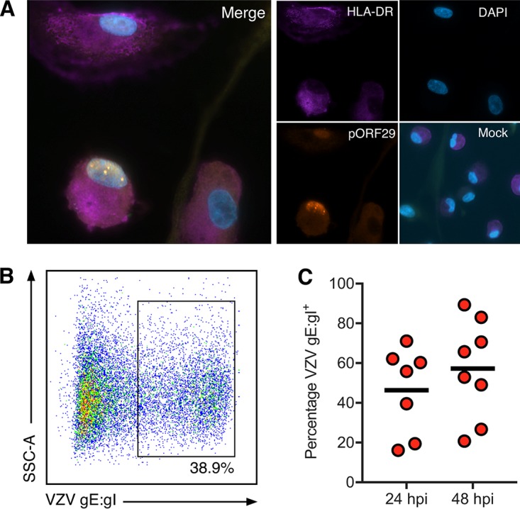
Varicella-zoster virus (VZV) is associated with viremia during primary infection that is presumed to stem from infection of circulating immune cells. While VZV has been shown to be capable of infecting a number of different subsets of circulating immune cells, such as T cells, dendritic cells, and NK cells, less is known about the interaction between VZV and monocytes. Here, we demonstrate that blood-derived human monocytes are permissive to VZV replication in vitro VZV-infected monocytes exhibited each temporal class of VZV gene expression, as evidenced by immunofluorescent staining. VZV virions were observed on the cell surface and viral nucleocapsids were observed in the nucleus of VZV-infected monocytes by scanning electron microscopy. In addition, VZV-infected monocytes were able to transfer infectious virus to human fibroblasts. Infected monocytes displayed impaired dextran-mediated endocytosis, and cell surface immunophenotyping revealed the downregulation of CD14, HLA-DR, CD11b, and the macrophage colony-stimulating factor (M-CSF) receptor. Analysis of the impact of VZV infection on M-CSF-stimulated monocyte-to-macrophage differentiation demonstrated the loss of cell viability, indicating that VZV-infected monocytes were unable to differentiate into viable macrophages. In contrast, macrophages differentiated from monocytes prior to exposure to VZV were highly permissive to infection. This study defines the permissiveness of these myeloid cell types to productive VZV infection and identifies the functional impairment of VZV-infected monocytes.IMPORTANCE Primary VZV infection results in the widespread dissemination of the virus throughout the host. Viral transportation is known to be directly influenced by susceptible immune cells in the circulation. Moreover, infection of immune cells by VZV results in attenuation of the antiviral mechanisms used to control infection and limit spread. Here, we provide evidence that human monocytes, which are highly abundant in the circulation, are permissive to productive VZV infection. Furthermore, monocyte-derived macrophages were also highly permissive to VZV infection, although VZV-infected monocytes were unable to differentiate into macrophages. Exploring the relationships between VZV and permissive immune cells, such as human monocytes and macrophages, elucidates novel immune evasion strategies and provides further insight into the control that VZV has over the immune system.

Keywords: varicella-zoster virus.


Figures

FIG 1
FIG 1
Isolation and infection of human CD14+ monocytes by VZV. (A) Preisolation PBMC and the positive fraction of anti-CD14 microbead isolation. The boxes display the proportion of live and single-cell monocytes expressing CD14 and CD11b, and the results are representative of those for 33 biologically independent donors. (B) CD14+ monocytes were cultured with mock- or VZV-infected HFF and assessed after 24, 48, and 72 h of culture. Representative plots indicate expression of VZV gE:gI on mock-infected (left) and VZV-infected (right) monocytes. Gating was performed on live, single cells, to the exclusion of the CFSE-labeled HFF inoculum. SSC-A, side scatter area. (C) VZV-infected monocytes exhibited cell surface VZV gE:gI+ at 24 hpi (n = 33), 48 hpi (n = 29), and 72 hpi (n = 5). The bars indicate the mean proportion at each time point.
FIG 2
FIG 2
VZV-infected monocytes display hallmarks of productive viral infection. VZV-infected monocytes were examined at 48 hpi by immunofluorescence staining for the presence and localization of immediate early protein IE62, early protein pORF29, and late glycoprotein gE (red staining) and DAPI nuclei (blue staining). Representative images of merged channels and individual viral antigen staining, mock-infected monocytes, and isotype control antibody-stained VZV-infected cells are shown. All images were acquired at a ×63 magnification and are representative of those from 5 independent experiments.
FIG 3
FIG 3
Backscattered scanning electron microscopy of VZV-infected human monocytes. VZV-infected monocytes were collected and sorted by FACS. VZV gE:gI+ monocytes were fixed and processed for serial block-face backscattered scanning electron microscopy. (A) Images depict a representative VZV gE:gI+ monocyte, with the two insets (i and ii) being magnified on the right. Arrows, VZV virions; arrowheads, virions with observable capsid structures. (B) Images depict a representative VZV gE:gI+ monocyte, with the inset, magnified on the right, showing VZV capsid structures within the nucleus (open arrowheads).
FIG 4
FIG 4
Transfer of infectious virus to HFF. (A) VZV-infected monocytes were isolated by FACS and seeded onto uninfected HFF monolayers for 5 days. The results of immunofluorescence assay (IFA) staining for early VZV pORF29 and late VZV gE shows infectious centers are presented. A merged IFA image of VZV-infected monocytes inoculated on HFF monolayers is shown. Individual channels show pORF29 (green), gE (red), DAPI (blue), and a merged image of mock-infected monocyte inoculation. All images were taken at a ×20 magnification and are representative of those from 3 independent experiments. (B) VZV-infected monocytes were pretreated with citrate buffer prior to inoculation onto uninfected HFF monolayers. A merged IFA image of citrate buffer-treated VZV-infected monocytes inoculated on HFF monolayers is shown. Individual channels of VZV gE (red) and DAPI (blue) are depicted. All images were taken at a ×20 magnification and are representative of those from 3 independent experiments. (C) Enumeration of infectious centers observed following inoculation of untreated and citrate buffer-treated, VZV-infected monocytes onto uninfected HFF. Each symbol represents an independent donor.
FIG 5
FIG 5
Viability of VZV-infected monocytes. Monocytes without exposure to HFF and mock- and VZV-infected monocytes were stained with live/dead dye (L/D) and antibodies for VZV gE:gI and intracellular cleaved caspase-3 (CC3). (A) VZV-infected monocytes were identified as VZV gE:gI+ (red box), and VZV-exposed monocytes were identified as VZV gE:gI (orange box). (B) Representative plot of VZV-infected monocytes stratified into viable cells (CC3, L/D) and cells with early apoptotic (CC3+, L/D), late apoptotic (CC3+, L/D+), and nonapoptotic (CC3, L/D+) cell death. (C) The proportions of monocytes (±SEM) at 24 hpi (top) and 48 hpi (bottom) are shown. Statistics were performed on the proportions of cells by repeated-measures two-way analysis of variance with Tukey’s multiple-comparison test. *, P < 0.05; **, P < 0.005; ***, P < 0.0005; ****, P < 0.00005; no symbol, an insignificant change.
FIG 6
FIG 6
VZV-infected monocytes exhibit a decreased endocytic ability. Mock- and VZV-infected monocytes at 24 hpi were incubated with 1 mg/ml dextran-FITC for 1 h at 37°C or 4°C. Flow cytometry gating was performed on live, single cells, prior to assessment of VZV infection by VZV gE:gI staining. (A) Representative histogram of dextran-FITC (Dx) uptake at 37°C and 4°C and monocytes without dextran (filled). (B) The MFI of surface-bound dextran-FITC at 4°C subtracted from the MFI of surface and internalized dextran-FITC at 37°C to generate the internalized MFI (±SEM). The internalized MFI of VZV-infected monocytes was normalized (norm.) to that of the respective mock-infected monocytes in 4 independent experiments. Statistical analyses were performed by paired two-tailed Student's t test. ****, P < 0.00005.
FIG 7
FIG 7
Cell surface immunophenotyping of VZV-infected monocytes. (A) Mock-infected monocytes (blue) and VZV-infected (red) monocytes were examined for the proportions of cells expressing the indicated cell surface markers at 24 and 48 hpi by flow cytometry. Gating was performed on live, single cells, prior to assessment of VZV infection by VZV gE:gI staining. Symbols depict the proportions of monocytes from multiple independent donors expressing the indicated markers, and bars represent the mean. (B) The MFI of immune markers on VZV-infected monocytes was normalized to that of the respective mock-infected monocytes at each time point. Bars represent the mean MFI (±SEM). Results are from multiple independent experiments examining CD11b (n = 8), HLA-ABC (n ≥ 8), HLA-DR (n ≥ 8), CD14 (n ≥ 18), and M-CSFR (n ≥ 9). Statistical analyses were performed for proportional and MFI analysis by paired two-tailed Student's t test. *, P < 0.05; **, P < 0.005; ***, P < 0.0005; ****, P < 0.00005; ns, not significant.
FIG 8
FIG 8
Differentiation of VZV-infected monocytes. Mock- and VZV-infected monocytes were collected at 24 hpi (day 0) and adhered into tissue culture plates in serum-free medium for 2 h at 37°C under 5% CO2. Nonadherent monocytes were aspirated, and the cultures were maintained for 3 days in medium supplemented with 25 ng/ml M-CSF (Peprotech). Adherent monocytes were collected by gentle scraping into the medium, stained for viability and VZV gE:gI, and analyzed by flow cytometry. (A) Timeline for infection and seeding. (B) Proportions of viable adherent monocytes cultured with mock-infected (blue) or VZV-infected (red) HFF. (C) Proportions of VZV gE:gI+ monocytes across each day measured for four independent donors.
FIG 9
FIG 9
VZV infection of monocyte-derived macrophages. Freshly isolated monocytes were adhered under serum-free conditions as described in the text and cultured in 25 ng/ml M-CSF for 6 days to generate macrophages (M-Mφ). M-CSF macrophages were cocultured with VZV-infected CFSE-labeled HFF for 24 and 48 h. VZV exposed M-Mφ were examined by IFA for the presence of HLA-DR and VZV pOR29. (A) Merged IFA image of VZV-infected M-Mφ at 48 hpi. Individual channels of HLA-DR (purple), pORF29 (orange), and DAPI (blue) are shown. A merged image of mock-infected M-Mφ at the same time point is also shown. All images were taken at a ×20 magnification and are representative of those from 4 independent experiments. Macrophages were collected at 24 and 48 hpi and assessed for VZV gE:gI by flow cytometry. Gating was performed on live, single cells and excluded the CFSE-labeled HFF inoculum. (B) VZV-infected M-Mφ exhibited cell surface VZV gE:gI. (C) VZV-infected M-Mφ displayed surface VZV gE:gI at 24 hpi (n = 4) and 48 hpi (n = 5). Bars indicate the mean proportion at each time point.

Similar articles

Cited by

References

    1. Arvin A. 2001. Varicella-zoster virus In Knipe DM, Howley PM, Griffin DE, Lamb RA, Martin MA, Roizman B, Straus SE (ed), Fields virology, 4th ed Lippincott Williams & Wilkins, Philadelphia, PA.
    1. Lungu O, Annunziato PW, Gershon A, Staugaitis SM, Josefson D, LaRussa P, Silverstein SJ. 1995. Reactivated and latent varicella-zoster virus in human dorsal root ganglia. Proc Natl Acad Sci U S A 92:10980–10984. doi:10.1073/pnas.92.24.10980. - DOI - PMC - PubMed
    1. Mainka C, Fuss B, Geiger H, Hofelmayr H, Wolff MH. 1998. Characterization of viremia at different stages of varicella-zoster virus infection. J Med Virol 56:91–98. doi:10.1002/(SICI)1096-9071(199809)56:1<91::AID-JMV15>3.0.CO;2-Z. - DOI - PubMed
    1. Satyaprakash AK, Tremaine AM, Stelter AA, Creed R, Ravanfar P, Mendoza N, Mehta SK, Rady PL, Pierson DL, Tyring SK. 2009. Viremia in acute herpes zoster. J Infect Dis 200:26–32. doi:10.1086/599381. - DOI - PubMed
    1. Vafai A, Wellish M, Gilden DH. 1988. Expression of varicella-zoster virus in blood mononuclear cells of patients with postherpetic neuralgia. Proc Natl Acad Sci U S A 85:2767–2770. doi:10.1073/pnas.85.8.2767. - DOI - PMC - PubMed

Publication types

MeSH terms

Substances