. 2019 Jan 17;93(3):e01375-18.
doi: 10.1128/JVI.01375-18. Print 2019 Feb 1.

Reactivation of Simian Varicella Virus in Rhesus Macaques after CD4 T Cell Depletion

Affiliations

Reactivation of Simian Varicella Virus in Rhesus Macaques after CD4 T Cell Depletion

Vicki Traina-Dorge et al. J Virol. .

Abstract

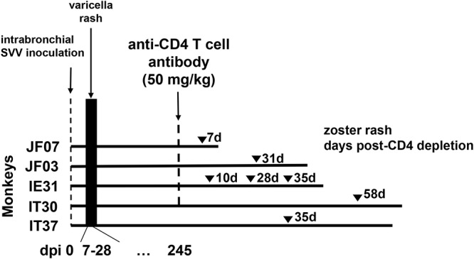
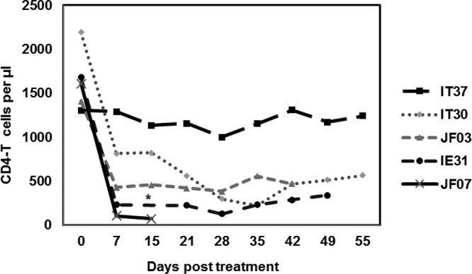
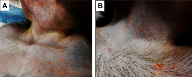
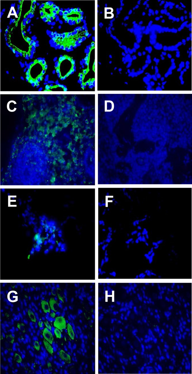
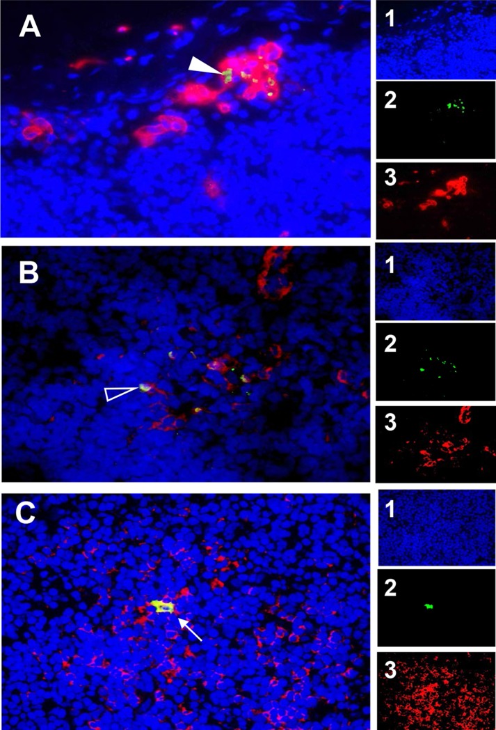
Rhesus macaques intrabronchially inoculated with simian varicella virus (SVV), the counterpart of human varicella-zoster virus (VZV), developed primary infection with viremia and rash, which resolved upon clearance of viremia, followed by the establishment of latency. To assess the role of CD4 T cell immunity in reactivation, monkeys were treated with a single 50-mg/kg dose of a humanized monoclonal anti-CD4 antibody; within 1 week, circulating CD4 T cells were reduced from 40 to 60% to 5 to 30% of the total T cell population and remained low for 2 months. Very low viremia was seen only in some of the treated monkeys. Zoster rash developed after 7 days in the monkey with the most extensive CD4 T cell depletion (5%) and in all other monkeys at 10 to 49 days posttreatment, with recurrent zoster in one treated monkey. SVV DNA was detected in the lung from two of five monkeys, in bronchial lymph nodes from one of the five monkeys, and in ganglia from at least two dermatomes in three of five monkeys. Immunofluorescence analysis of skin rash, lungs, lymph nodes, and ganglia revealed SVV ORF63 protein at the following sites: sweat glands in skin; type II cells in lung alveoli, macrophages, and dendritic cells in lymph nodes; and the neuronal cytoplasm of ganglia. Detection of SVV antigen in multiple tissues upon CD4 T cell depletion and virus reactivation suggests a critical role for CD4 T cell immunity in controlling varicella virus latency.IMPORTANCE Reactivation of latent VZV in humans can result in serious neurological complications. VZV-specific cell-mediated immunity is critical for the maintenance of latency. Similar to VZV in humans, SVV causes varicella in monkeys, establishes latency in ganglia, and reactivates to produce shingles. Here, we show that depletion of CD4 T cells in rhesus macaques results in SVV reactivation, with virus antigens found in zoster rash and SVV DNA and antigens found in lungs, lymph nodes, and ganglia. These results suggest the critical role of CD4 T cell immunity in controlling varicella virus latency.

Keywords: CD4 T cell depletion; animal model; simian varicella virus reactivation.


Figures

FIG 1
FIG 1
Experimental design. Five rhesus macaques (JF07, JF03, IE31, IT30, and IT37) were inoculated intrabronchially with SVV (105 PFU). All monkeys developed varicella rash 7 to 14 days after inoculation. Eight months (245 days) later, four of the monkeys (JF07, JF03, IE31, and IT30) were treated intravenously with 50 mg/kg of anti-CD4 antibody; monkey IT37 was left untreated. Seven days after CD4 depletion, monkey JF07 developed zoster rash. Monkey JF03 developed zoster 28 days after CD4 depletion, while monkey IE31 developed zoster 10, 28, and 35 days after CD4 depletion. Monkey IT30 developed zoster 49 days after CD4 depletion. Monkey IT37 developed zoster 38 days after the CD4 depletion treatment in the other 4 monkeys. Monkeys JF07, JF03, IE31, IT30, and IT37 were euthanized 9, 16, 15, 35, and 33 days, respectively, after the last episode of zoster, and tissues were collected for analysis.
FIG 2
FIG 2
Reduction in CD4 T lymphocyte levels after treatment of latently infected rhesus macaques with anti-CD4 antibodies. Blood samples collected between 0 and 55 days after CD4 depletion were used for analysis. Absolute numbers of CD4 T cells/µl in peripheral blood were plotted against the days posttreatment for all rhesus macaques latently infected with SVV. *, The sample from monkey IE31 at 15 dpix was not available.
FIG 3
FIG 3
Skin rash after varicella and zoster in the same monkey. Skin rash was seen in the upper thorax and chin 9 days after SVV inoculation (primary infection) (A) and in the upper thorax and neck 35 days after CD4 depletion (zoster) (B) in monkey IE31.
FIG 4
FIG 4
Detection of SVV antigens in tissues of monkeys after SVV reactivation after CD4 depletion. Paraformaldehyde-fixed, paraffin-embedded tissue sections were analyzed by immunofluorescence. (A) In monkey JF03, SVV ORF63 protein was found in the sweat glands in skin with zoster rash using rabbit polyclonal antibodies specific for SVV ORF63 peptides. SVV glycoproteins gH and gL were detected using rabbit polyclonal antibodies raised against peptides specific for these glycoproteins in the medulla of axillary lymph nodes from monkey IE31 (C), in the lung alveoli of monkey JF07 (E), and in the cytoplasm of neurons in thoracic ganglia from monkey JF07 (G). No staining was seen in the respective adjacent sections when preimmune rabbit serum was substituted for anti-SVV antibody (B, D, F, and H). Donkey Alexa Fluor 488-tagged anti-rabbit IgG(H+L) antibody was used as a secondary antibody and visualized at 385 nm. DAPI (4′,6′-diamidino-2-phenylindole) staining shows the nuclei of cells. Magnification, ×600.
FIG 5
FIG 5
Detection of SVV ORF63 protein in macrophages, dendritic cells, and CD4 T cells in the lymph nodes of rhesus macaques. Paraformaldehyde-fixed, paraffin-embedded sections of axillary (A) and bronchial (B) lymph nodes from monkey IE31 and in axillary lymph node from monkey IT37 (C) were analyzed by immunofluorescence using rabbit polyclonal antibody specific for SVV ORF63 protein and mouse anti-human CD163 (macrophage) antibody (A), mouse anti-human CD123 (dendritic cells) antibody (B), or mouse anti-human CD4 T cell antibody (C). Donkey Alexa Fluor 488-tagged anti-rabbit IgG(H+L) antibody and goat Alexa Fluor 594-tagged anti-mouse IgG(H+L) antibody were used as secondary antibodies. Nuclei were stained with DAPI and visualized at 358 nm. The colocalization of SVV IE63 in macrophages (filled arrowhead), dendritic cells (open arrowhead), and CD4 T cells (arrow) is indicated. Images of individual channels are presented in panels A1 to 3, B1 to 3, and C1 to 3. Magnification, ×600.
FIG 6
FIG 6
Detection of SVV ORF63 protein in epithelial type II cells within the alveolar surface. Paraformaldehyde-fixed, paraffin-embedded sections of lung from monkey JF07 were analyzed by immunofluorescence using rabbit polyclonal antibody specific for SVV IE63 protein and mouse monoclonal antibody specific for ABCA3 protein. SVV ORF63 protein colocalized with ABCA3 protein in the alveolar surface (arrowhead). The fluorescence-tagged secondary antibodies are as described in the legend to Fig. 5. DAPI staining shows the nuclei of cells. Images of individual channels are presented in panels 1 to 3. Magnification, ×600.

Similar articles

Cited by

References

    1. Levin MJ, Smith JG, Kaufhold RM, Barber D, Hayward AR, Chan CY, Chan IS, Li DJ, Wang W, Keller PM, Shaw A, Silber JL, Schlienger K, Chalikonda I, Vessey SJ, Caulfield MJ. 2003. Decline in varicella-zoster virus (VZV)-specific cell-mediated immunity with increasing age and boosting with a high-dose VZV vaccine. J Infect Dis 188:1336–1344. doi:10.1086/379048. - DOI - PubMed
    1. Hess TM, Lutz LJ, Nauss LA, Lamer TJ. 1990. Treatment of acute herpetic neuralgia: a case report and review of the literature. Minn Med 73:37–40. - PubMed
    1. Nagel MA. 2014. Varicella zoster virus vasculopathy: clinical features and pathogenesis. J Neurovirol 20:157–163. doi:10.1007/s13365-013-0183-9. - DOI - PMC - PubMed
    1. Matoba AY, Meghpara B, Chevez-Barrios P. 2014. Varicella-zoster virus detection in varicella-associated stromal keratitis. JAMA Ophthalmol 132:505–506. doi:10.1001/jamaophthalmol.2013.8254. - DOI - PubMed
    1. Gilden D, Nagel MA. 2016. Varicella zoster virus triggers the immunopathology of giant cell arteritis. Curr Opin Rheumatol 28:376–382. doi:10.1097/BOR.0000000000000292. - DOI - PMC - PubMed

Publication types

MeSH terms