. 2019 Jan 17;93(3):e01803-18.
doi: 10.1128/JVI.01803-18. Print 2019 Feb 1.

Herpes Simplex Virus 1 ICP22 Suppresses CD80 Expression by Murine Dendritic Cells

Affiliations

Herpes Simplex Virus 1 ICP22 Suppresses CD80 Expression by Murine Dendritic Cells

Harry Matundan et al. J Virol. .

Abstract

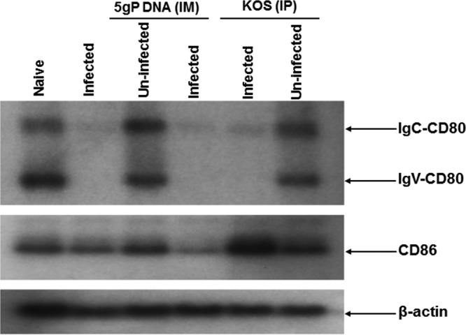
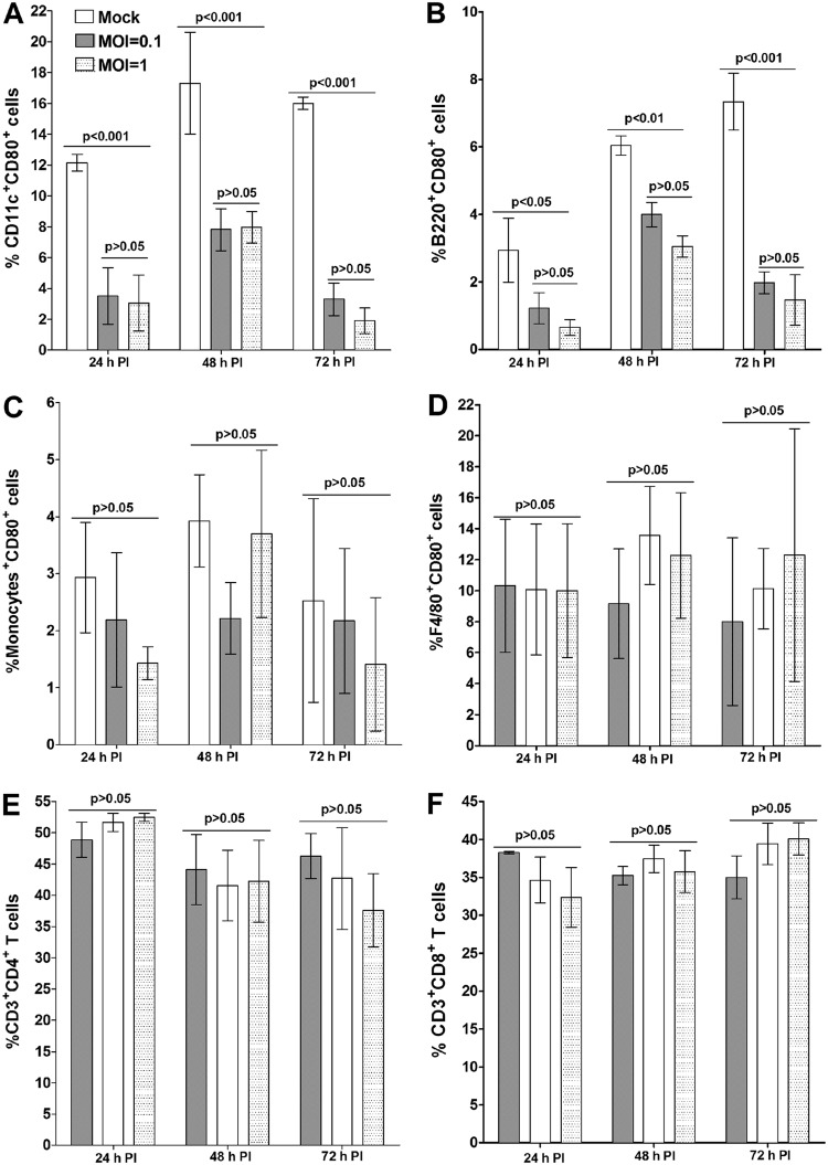
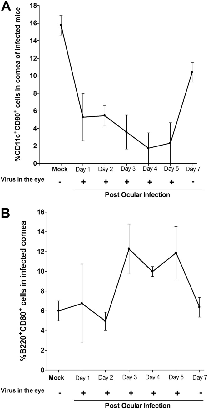
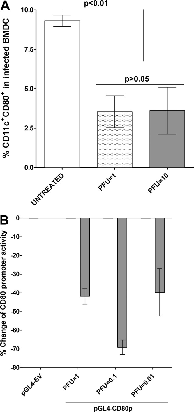
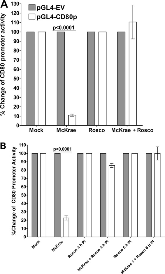
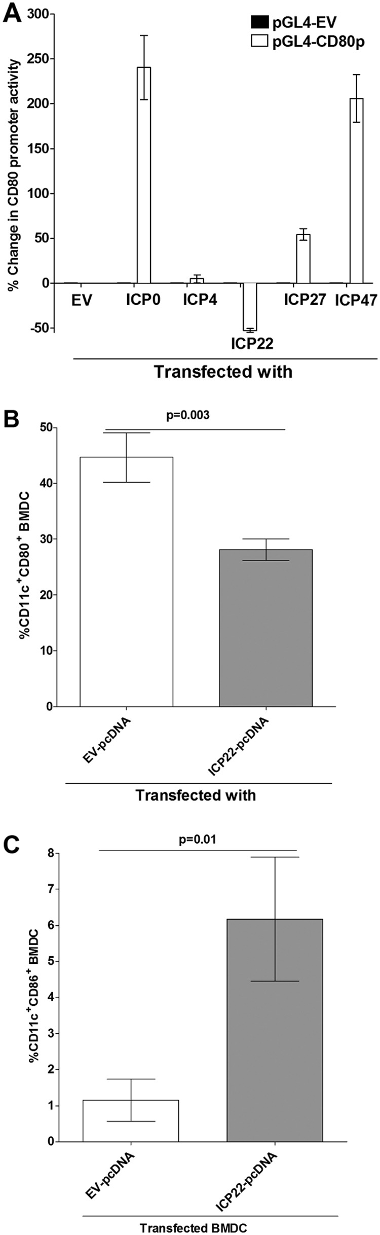
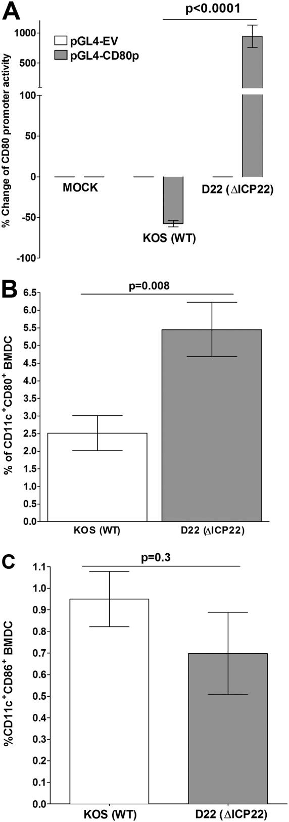
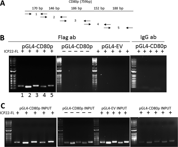
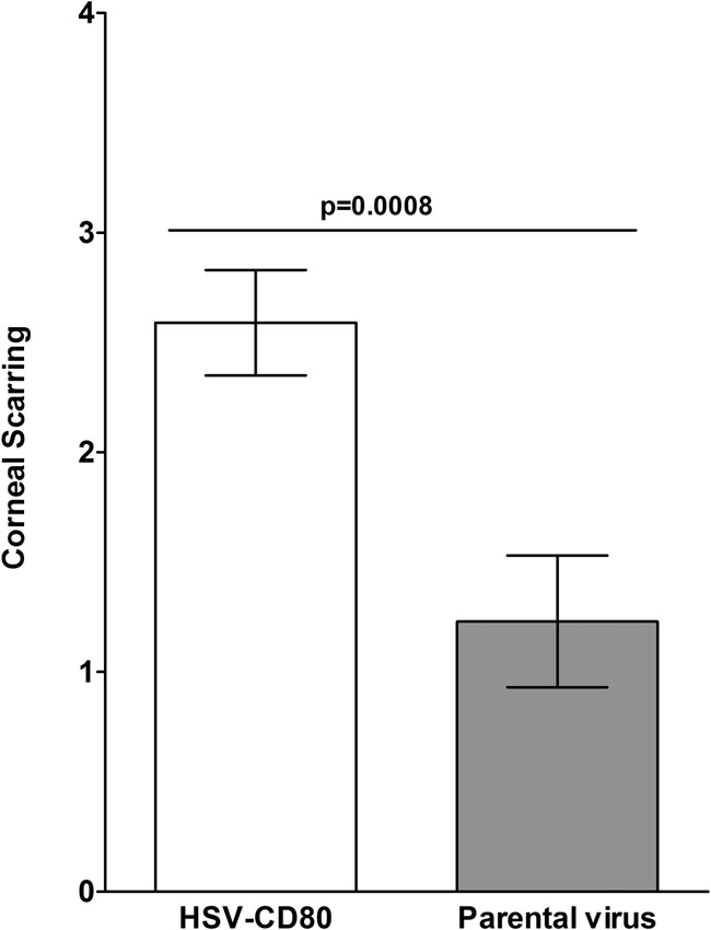
Herpes simplex virus type 1 (HSV-1) has the ability to delay its clearance from the eye during ocular infection. Here, we show that ocular infection of mice with HSV-1 suppressed expression of the costimulatory molecule CD80 but not CD86 in the cornea. The presence of neutralizing anti-HSV-1 antibodies did not alleviate this suppression. At the cellular level, HSV-1 consistently downregulated the expression of CD80 by dendritic cells (DCs) but not by other antigen-presenting cells. Furthermore, flow cytometric analysis of HSV-1-infected corneal cells during a 7-day period reduced CD80 expression in DCs but not in B cells, macrophages, or monocytes. This suppression was associated with the presence of virus. Similar results were obtained using infected or transfected spleen cells or bone marrow-derived DCs. A combination of roscovitine treatment, transfection with immediate early genes (IE), and infection with a recombinant HSV-1 lacking the ICP22 gene shows the importance of ICP22 in downregulation of the CD80 promoter but not the CD86 promoter in vitro and in vivo At the mechanistic level, we show that the HSV-1 immediate early gene ICP22 binds the CD80 promoter and that this interaction is required for HSV-1-mediated suppression of CD80 expression. Conversely, forced expression of CD80 by ocular infection of mice with a recombinant HSV-1 exacerbated corneal scarring in infected mice. Taken together, these studies identify ICP22-mediated suppression of CD80 expression in dendritic cells as central to delayed clearance of the virus and limitation of the cytopathological response to primary infection in the eye.IMPORTANCE HSV-1-induced eye disease is a major public health problem. Eye disease is associated closely with immune responses to the virus and is exacerbated by delayed clearance of the primary infection. The immune system relies on antigen-presenting cells of the innate immune system to activate the T cell response. We found that HSV-1 utilizes a robust and finely targeted mechanism of local immune evasion. It downregulates the expression of the costimulatory molecule CD80 but not CD86 on resident dendritic cells irrespective of the presence of anti-HSV-1 antibodies. The effect is mediated by direct binding of HSV-1 ICP22, the product of an immediate early gene of HSV-1, to the promoter of CD80. This immune evasion mechanism dampens the host immune response and, thus, reduces eye disease in ocularly infected mice. Therefore, ICP22 may be a novel inhibitor of CD80 that could be used to modulate the immune response.

Keywords: antigen-presenting cells; cornea; corneal scarring; eye disease; immediate early genes; immune suppression; virus replication.


Figures

FIG 1
FIG 1
Southern analyses. BALB/c mice were inoculated with DNA of five glycoproteins (gB, gC, gD, gE, and gI [5gP]) or avirulent HSV-1 strain KOS or mock treated as described in Materials and Methods. Inoculated or mock-treated mice were ocularly infected with 2 × 105 PFU/eye of virulent HSV-1 strain McKrae virus. As a control, some of the inoculated or mock-treated mice were not ocularly infected. Corneas from 3 mice per treatment were isolated at 5 days p.i. and combined, and total RNA was extracted. cDNA synthesis was performed on the total extracted RNA, and the cDNAs were separated using a 0.9% agarose gel, transferred to Zeta paper, rinsed in 2 × SSC (1 × SSC is 0.15 M NaCl plus 0.015 M sodium citrate) for 5 min, and cross-linked to the membrane by UV light. DNA-DNA hybridization was carried out using 32P-labeled CD80, CD86, or the β-actin gene (as a control) as we described previously (85).
FIG 2
FIG 2
Suppression of CD80 expression by DCs in HSV-1 infected splenocytes in vitro. Cultured splenocytes from naive mice were infected with 0.1 or 1 PFU/cell of HSV-1 strain McKrae or mock infected. Infected or mock-infected cells were collected at 24, 48, and 72 h p.i. for FACS analysis as described in Materials and Methods. Cells were stained with anti-CD11c, anti-CD11b, anti-B220, anti-F4/80, anti-CD3, anti-CD4, anti-CD8, and anti-CD80 antibodies. FACS analysis was performed by gating for CD11c+ CD80+ cells, CD11b+ CD80+ cells, B220+ CD80+ cells, F4/80+ CD80+ cells, CD3+ CD4+ CD80+ cells, and CD3+ CD8+ CD80+ cells, as indicated. Data are presented as means ± SEM from three independent studies and with three replications per each experiment (n = 9). Statistical analysis was done using two-way ANOVA to test for P values.
FIG 3
FIG 3
HSV-1 infection suppresses CD80 expression in cornea of infected mice. C57BL/6 mice were infected in both eyes with 2 × 105 PFU/eye of HSV-1 strain McKrae. Corneas from infected mice were isolated on days 1, 2, 3, 4, 5, and 7 p.i. Isolated corneas from each mouse were digested with collagenase and washed, and the cell suspension was stained with anti-CD11c, anti-B220, and anti-CD80 antibodies prior to flow cytometry analysis as described in Materials and Methods. Tear films were also collected on the indicated days, and virus titers were determined using standard plaque assays. Quantitation of the mean number of CD11c+ CD80+ cells or B220+ CD80+ cells ± SEM from three independent experiments with three replications per individual mouse corneas is shown (n = 9). The presence (+) or the absence (−) of virus in tear films of infected mice for each indicated time point is shown. Each point represents the mean ± SEM for 10 mice (20 eyes) per time point.
FIG 4
FIG 4
Suppression of CD80 in infected BMDCs. Subconfluent monolayers of DCs isolated from C57BL/6 mice were infected with 1 or 10 PFU/cell of HSV-1 strain McKrae for 24 h as described in Materials and Methods. In some experiments DCs were transfected with pGL4-CD80p or pGL4-EV and 72 h later infected with 1, 0.1, or 0.01 PFU/cell of McKrae virus for 24 h. Untreated DCs were used as a control for both groups. In the first group, infected DCs were stained with anti-CD11c and anti-CD80 antibodies, and percentages of CD11c+ CD80+ cells ± SEM from three independent experiments with three replications of each experiment are shown (n = 9). In the transfected and infected DCs, the changes in CD80 promoter activity were determined as described in Materials and Methods. Each point represents the mean ± SEM (n = 9) from three separate experiments. Statistical analyses were done using ANOVA to test for P values.
FIG 5
FIG 5
Effects of roscovitine treatment on CD80 promoter activity. HEK 293 cells were grown and transfected with pGL4-EV or pGL4-CD80p DNA as described in the legend of Fig. 4. Transfected cells were incubated with McKrae virus, roscovitine, or both, and CD80 promoter activity was determined 1 h after roscovitine treatment (A) as described in Materials and Methods. In some of the experiments, roscovitine treatment was carried out at 4 h and 6 h p.i. instead of at 0 h p.i. (B). Each point represents the mean ± SEM from three separate experiments (n = 9). Statistical analyses were done using ANOVA to test for P values.
FIG 6
FIG 6
Effects of immediate early genes on CD80 promoter activity. (A) Effect of IE genes on transfected HEK 293 cells. HEK 293 cells were transfected with either pGL4-EV or pGL4-CD80p DNA and 24 h later were individually transfected with ICP0, ICP4, ICP22, ICP27, or ICP47 DNA, and their effect on CD80 promoter activity was determined 48 h later as described in Materials and Methods. (B and C) Effect of ICP22 on BMDCs. BMDCs were transfected with either EV-pcDNA or ICP22-pcDNA for 24 h as described in Materials and Methods. Transfected cells were stained with anti-CD11c and anti-CD80 or with anti-CD11c and anti-CD86 antibodies, and the effects of ICP22 on CD80 (B) and CD86 (C) expression were determined by flow cytometric analysis. Each point represents the mean ± SEM from three separate experiments (n = 9). Statistical analyses were done using ANOVA to test for P values.
FIG 7
FIG 7
Effects of an ICP22 deletion mutant HSV-1 virus on CD80 expression. (A) Effects of ICP22 deletion virus in transfected HEK 293 cells. HEK 293 cells were transfected with either pGL4-EV or pGL4-CD80p DNA and 24 h later were infected with 10 PFU/cell of D22 (ΔICP22) virus that lacks the ICP22 gene or its KOS parental virus or were mock infected for 24 h. CD80 promoter activity was determined at 24 h p.i. as described in Materials and Methods. (B and C) Effect of ICP22 deletion virus in BMDCs. BMDCs were infected with 10 PFU/cell of D22 or KOS as described for panel A for 24 h. At 24 h p.i., infected BMDCs were stained with anti-CD11c and anti-CD80 antibodies or anti-CD11c and anti-CD80 antibodies, and the effects of ICP22 deletion on CD80 (B) and CD86 (C) were determined by flow cytometric analysis. Each point represents the mean ± SEM from three separate experiments with three replications (n = 9). Statistical analyses were done using ANOVA to test for P values.
FIG 8
FIG 8
Chromatin immunoprecipitation (ChIP) of ICP22-bound chromatin. (A) Schematic of the primers pairs used for PCR of the five sections of the CD80 promoter. Primer pairs are designated with arrows that represent forward and reverse primers with corresponding expected product sizes, which are positioned over the corresponding region (e.g., primer pair 1 has a size of 170 bp). (B) Chromatin immunoprecipitation and PCR amplification of CD80 promoter. Subconfluent HEK 293 cells were transfected with either pGL4-CD80p or pGL4-EV and pCDNA3.1-ICP22-FLAG (FL), in combinations as described in Materials and Methods. Transfected cells were harvested and lysed, and the lysate was sheared by sonication and prepared for pulldown. Protein G magnetic beads were incubated with anti-FLAG antibody, and the lysates were incubated overnight at 4°C. A stationary magnet was used to immobilize the chromatin-FLAG-MAb-protein G magnetic bead complex, and subsequent elution, reverse cross-linking, and proteinase K completed the preparation of the chromatin template. The PCR products were loaded on a 1% agarose gel along with a 1-kb Plus ladder (Invitrogen). The FLAG antibody pulldown of samples was transfected using the following plasmids: pGL4-CD80p and pCDNA3.1-ICP22-FL, pGL4-CD80p alone, and pGL4-EV with pCDNA3.1-ICP22-FL. An isotype IgG control pulldown with pGL4-CD80p and pCDNA3.1-ICP22-FL was included. (C) PCR experiments of input chromatin. The PCR experiment utilized chromatin templates that did not undergo FLAG pulldown but otherwise were similar with regard to primers used and transfection combinations, as described above. The putative binding site for ICP22 on the CD80 promoter falls between positions 151 and 462 of the CD80 promoter, and the total fragment size for the two primer sets involved (set 2 and set 3), which includes the primers, is 311 bp (the CD80 promoter sequence is shown in bold in Fig. S1 in the supplemental material).
FIG 9
FIG 9
Corneal scarring in mice infected ocularly with HSV-CD80. BALB/c mice were ocularly infected with 1 × 105 PFU/eye of HSV-CD80 or its parental virus. Corneal scarring (CS) in surviving mice was examined on day 28 p.i. as described in Materials and Methods. The CS score represents the average ± SEM from 34 and 26 eyes for HSV-CD80 and parental virus infection, respectively.

Similar articles

Cited by

References

    1. Barron BA, Gee L, Hauck WW, Kurinij N, Dawson CR, Jones DB, Wilhelmus KR, Kaufman HE, Sugar J, Hyndiuk RA, Laibson PR, Stulting RD, Asbell PA, for the Herpetic Eye Disease Study Group. 1994. Herpetic eye disease study. A controlled trial of oral acyclovir for herpes simplex stromal keratitis. Ophthalmology 101:1871–1882 doi:10.1016/S0161-6420(13)31155-5. - DOI - PubMed
    1. Wilhelmus KR, Dawson CR, Barron BA, Bacchetti P, Gee L, Jones DB, Kaufman HE, Sugar J, Hyndiuk RA, Laibson PR, Stulting RD, Asbell PA. 1996. Risk factors for herpes simplex virus epithelial keratitis recurring during treatment of stromal keratitis or iridocyclitis. Herpetic Eye Disease Study Group. Br J Ophthalmol 80:969–972. doi:10.1136/bjo.80.11.969. - DOI - PMC - PubMed
    1. Liesegang TJ. 1999. Classification of herpes simplex virus keratitis and anterior uveitis. Cornea 18:127–143. doi:10.1097/00003226-199903000-00001. - DOI - PubMed
    1. Liesegang TJ. 2001. Herpes simplex virus epidemiology and ocular importance. Cornea 20:1–13. doi:10.1097/00003226-200101000-00001. - DOI - PubMed
    1. Hill TJ. 1987. Ocular pathogenicity of herpes simplex virus. Curr Eye Res 6:1–7. doi:10.3109/02713688709020060. - DOI - PubMed

Publication types

MeSH terms