. 2018 Nov 20;131(22):jcs217240.
doi: 10.1242/jcs.217240.

Fibronectin regulates growth factor signaling and cell differentiation in primary lens cells

Affiliations

Fibronectin regulates growth factor signaling and cell differentiation in primary lens cells

Judy K VanSlyke et al. J Cell Sci. .

Abstract

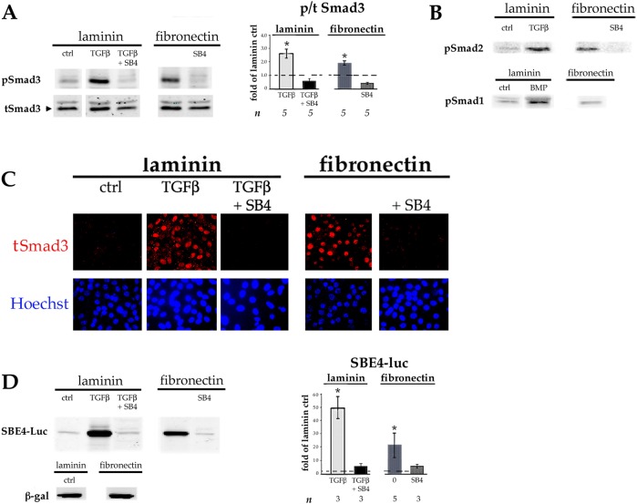
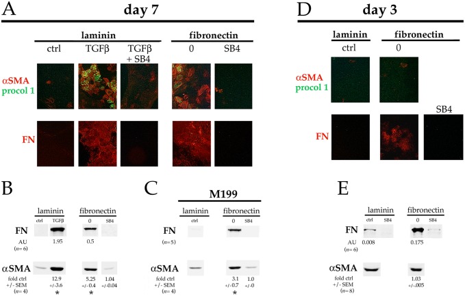
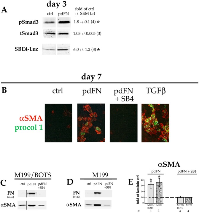
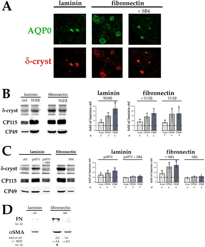
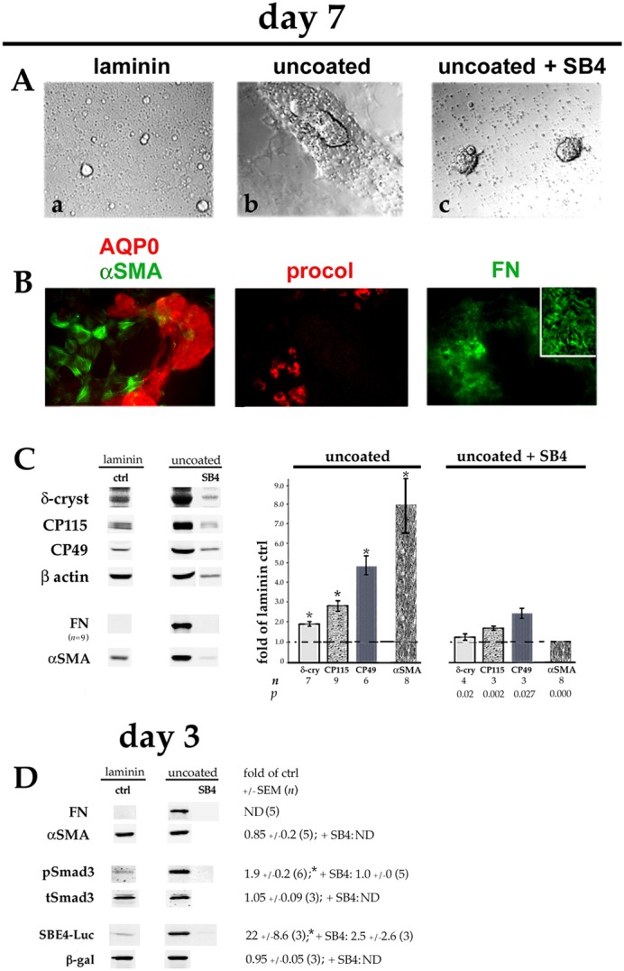
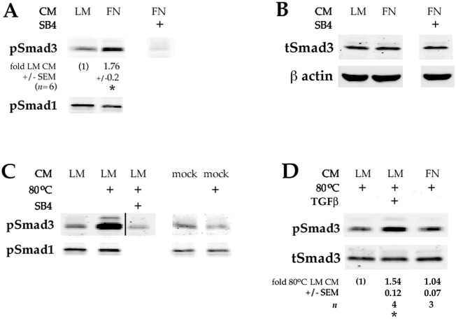
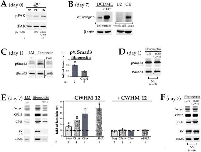
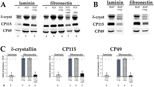
Lens epithelial cells are bound to the lens extracellular matrix capsule, of which laminin is a major component. After cataract surgery, surviving lens epithelial cells are exposed to increased levels of fibronectin, and so we addressed whether fibronectin influences lens cell fate, using DCDML cells as a serum-free primary lens epithelial cell culture system. We found that culturing DCDMLs with plasma-derived fibronectin upregulated canonical TGFβ signaling relative to cells plated on laminin. Fibronectin-exposed cultures also showed increased TGFβ signaling-dependent differentiation into the two cell types responsible for posterior capsule opacification after cataract surgery, namely myofibroblasts and lens fiber cells. Increased TGFβ activity could be identified in the conditioned medium recovered from cells grown on fibronectin. Other experiments showed that plating DCDMLs on fibronectin overcomes the need for BMP in fibroblast growth factor (FGF)-induced lens fiber cell differentiation, a requirement that is restored when endogenous TGFβ signaling is inhibited. These results demonstrate how the TGFβ-fibronectin axis can profoundly affect lens cell fate. This axis represents a novel target for prevention of late-onset posterior capsule opacification, a common but currently intractable complication of cataract surgery.

Keywords: Cataract; Epithelial-mesenchymal transition; Fibrosis; Lens; TGFβ.


Conflict of interest statement

Competing interestsThe authors declare no competing or financial interests.

Figures

Fig. 1.
Fig. 1.
Lens epithelial cells plated on FN have higher levels of canonical TGFβ signaling than cells plated on laminin. DCDMLs were plated on day 0 of culture in tissue culture wells coated with either laminin or pdFN. On day 1 of culture, the culture medium was replaced with fresh M199/BOTS with or without 4 ng/ml TGFβ1, the TGFβR1 inhibitor SB431542 (SB4), or TGFβ1 plus SB431542 as indicated. Cells were analyzed on day 3 of culture. (A,B) Whole-cell lysates were prepared and probed with antibodies specific for total Smad3 (tSmad3) or the phosphorylated (activated) forms of Smad3, Smad2 or Smad1 (pSmad1, pSmad2 or pSmad3) as indicated. (C) Cells were fixed and immunostained for total Smad3 and nuclei counterstained with Hoechst 33258. (D) DCDMLs were transfected with plasmids encoding either the SBE4–luciferase (SBE4–Luc) reporter construct or β-galactosidase (β-gal) on day 1 of culture prior to cell lysis on day 3. Expression of pSmad3 (A) and luciferase (D) as assessed using western blot analysis was normalized to β-actin in the same sample, and plotted as fold-change of expression versus control (ctrl; defined as DCDMLs plated on laminin and cultured in unsupplemented M199/BOTS). *P≤0.02; other data sets not significantly different from ctrl. B and C are representative of ≥3 experiments.
Fig. 2.
Fig. 2.
Plating on FN induces expression of fibrotic markers. DCDMLs plated on either laminin or pdFN on day 0 were cultured from day 1 to day 7 (A–C) or from day 1 to day 3 (D,E) with or without 4 ng/ml TGFβ1 and/or SB431542 (SB4). (A,D) Cultures were immunostained for the mesenchymal proteins procollagen 1 (procol 1) and fibronectin (FN), or the myofibroblast marker α smooth muscle actin (αSMA). Near-confluent regions of cultures are shown. All markers assessed in a minimum of three independent experiments with similar results. (B,C,E) Cells were assayed for expression of FN or αSMA using quantitative western blot. Cells in C were cultured in M199 medium without BOTS supplementation. In E, the amount of cell lysate analyzed was tripled, and western blots probed for FN and αSMA were scanned at increased intensity. Data for αSMA (normalized to β-actin in the same samples) was quantitated as fold-change versus the values obtained with cells plated on laminin and cultured without TGFβ or SB4 (ctrl) in the same experiment. *P≤0.009; all other data sets not significantly different from ctrl. Where expression was high enough to be reproducibly quantitated, FN levels were expressed as arbitrary units (AU) obtained using infrared imaging, normalized to the value obtained in the same sample for β-actin.
Fig. 3.
Fig. 3.
Adding pdFN to the culture medium induces TGFβ signaling and EMyT in lens cells. DCDMLs were plated on laminin on day 0 and cultured from day 1 in M199/BOTS (A–C) or M199 (D) medium with or without (ctrl) 12.5 µg/ml pdFN, or with pdFN and SB431542 (pdFN+SB4) as indicated. (A) Cultures were analyzed on day 3 for pSmad3, total Smad3 (tSmad3), or SBE4–luciferase (SBE4–Luc) expression as in Fig. 1. (B) Cells were fixed on day 7 prior to immunostaining for αSMA and procollagen 1 as in Fig. 2. The (negative) control image from Fig. 2A was repeated in Fig. 3B because these data were from the same experiment. (C,D) Cells were assayed on day 7 for expression of fibronectin (FN) or α smooth muscle actin (αSMA) using western blot as in Fig. 2. All data shown are from the same blot at the same exposure. (E) Data for αSMA (normalized to β-actin in the same samples) in C,D was quantitated as fold-change versus the values obtained with cells plated on laminin and cultured without TGFβ or SB4 (ctrl) in the same experiment. *P≤0.02; all other data sets, not significantly different from control (P>0.2).
Fig. 4.
Fig. 4.
FN also induces lens fiber cell differentiation. DCDMLs were plated on laminin or pdFN at 0.9×10⁵ cells/96 plate well on day 0 and cultured from day 1 in M199/BOTS (A,B,D) or M199 (C) medium with or without 4 ng/ml TGFβ1, 12.5 µg/ml pdFN, and/or SB431542 (SB4) as indicated. (A) On day 7 of culture, cells were fixed and double-immunostained for proteins specific to, or highly enriched in, differentiating lens fiber cells (aquaporin-0, AQP0; and δ-crystallin, respectively). Representative of ≥4 experiments. (B,C) On day 7 of culture, cells were assayed for synthesis of the fiber cell differentiation markers δ-crystallin (by [35S]-methionine labeling), CP115, and CP49 (by quantitative western blotting). The extent to which the treatment increased marker expression was calculated as fold-change versus the values obtained with cells plated on laminin and cultured with no additions (ctrl) in the same experiment. *P≤0.018; all other data sets, not significantly different than control (P>0.2). (D) Cells were assayed on day 7 for expression of the fibrotic markers FN and αSMA using quantitative western blot as in Fig. 2. *P=0.001.
Fig. 5.
Fig. 5.
Plating DCDMLs on uncoated tissue culture plastic upregulates EMyT, fiber cell differentiation and TGFβ signaling. DCDMLs were plated in uncoated tissue culture wells (or, as a control, in laminin-coated wells) at 1.2×10⁵ cells/96 plate well on day 0. Cells were cultured from day 1 in M199/BOTS with or without SB431542 (SB4) as indicated. (A) Phase-contrast images of cultures on day 7; (a,b) show near-confluent regions, whereas (c) shows two balls of tightly packed cells surrounded by phase-bright cell blebs. (B) Cultures plated on uncoated plastic were immunostained for aquaporin-0 (AQP0) and the fibrotic markers αSMA, procollagen 1 (procol), and FN. Near-confluent regions of cultures are shown; inset is a high-magnification view of NaOH-insoluble FN extracellular fibrils. All markers assessed in a minimum of three independent experiments with similar results. (C) Cells were assayed on day 7 for expression of fiber cell and EMT/EMyT markers as in Fig. 4. Note that the lower number of adherent cells in cultures plated on uncoated TC plastic and treated with SB431542 resulted in lower levels of β-actin per sample. (D) Cultures were analyzed on day 3 for FN and αSMA, or for pSmad3, total Smad3 (tSmad3) and SBE4–luciferase (SBE4–Luc) expression using quantitative western blot as in Fig. 1. *P≤0.001 compared to untreated cells plated on laminin (ctrl); all other data sets not significantly different from control (P>0.2) unless indicated otherwise. ND, not determined.
Fig. 6.
Fig. 6.
Plating lens cells on pdFN increases their ability to activate endogenous TGFβ. Conditioned medium donor cells were plated on laminin (LM) or plasma-derived fibronectin (FN) on day 0. Medium was replaced with fresh M199/BOTS on day 1, and the conditioned medium (CM) collected on day 3. The CM was then added to recipient DCDMLs plated on laminin and incubated for 1.5 h prior to lysis of recipient cells and western blot analysis of active (phosphorylated) Smad3 and Smad1 (pSmad3/1), or total Smad3 (tSmad3). Where indicated (80°C), CM was heated to 80°C for 6 min to thermally activate endogenous latent TGFβ prior to addition to recipient cells. In some cases, recipient cells were pretreated with SB431542 for 1 h prior to addition of CM. (A) CM from pdFN-plated cells has a ∼1.8-fold greater ability to enhance activation of Smad3 than CM from laminin-plated cells. *P<0.000 compared to unheated CM from laminin-plated cells. (B) Total Smad3 levels did not change during the course of the experiment. (C) Heat treatment of CM from laminin-plated cells increased the level of pSmad3 in recipient cells by 3.88-fold (±0.7; n=4; P=0.004). Mock CM was generated using a 48 h incubation of M199/BOTS in pdFN-coated wells without cells. pSmad1 levels did not change during the course of the experiment. All data shown are from the same blot at the same exposure. (D) Heated CM was diluted tenfold with fresh M199/BOTS before addition to recipient cells. This ensured that the pSmad3 signal was not saturated and reflected the level of TGFβ signaling, as demonstrated by the 1.54-fold increase (±0.12; n=4; P=0.003) in pSmad3 levels when 4 ng/ml (active) TGFβ was added to heated CM. The difference between the Smad3-activating activity of heated CM from donor cells plated on laminin or pdFN was not significant (P=0.465). Total Smad3 levels did not change during the course of the experiment.
Fig. 7.
Fig. 7.
FN-binding, TGFβ-activating integrins in DCDMLs. (A) Freshly prepared lens epithelial cells were left in suspension (sp), or plated on tissue culture wells coated with either poly-D-lysine (PL) or pdFN (FN). After 45 min, cells were lysed and analyzed using quantitative western blot for total FAK (tFAK) and, in the same sample, FAK autophosphorylated at Y397 (pFAK). (B) DCDMLs plated on either laminin or pdFN were cultured with or without TGFβ. On day 7, the cultures were subjected to cell-surface biotinylation at 4°C and assessed for α5 integrin on the plasma membrane using western blot. Fold-increase in cell-surface α5 integrin (normalized to β-actin in the corresponding whole cell lysate) in TGFβ-treated cells was 6.75×±0.2 (n=3; P=0.001). Equal amounts of total cell lysate from α5 integrin-deficient CHO-B2 cells (B2) (Wu et al., 1993) and α5 integrin-rich HH stage 17 chick embryos (CE) (Muschler and Horowitz, 1991) were used as negative and positive controls, respectively. (C) DCDMLs were plated on day 0 on either laminin (LM) or pdFN. After 4 h, the medium was replaced with fresh M199/BOTS with or without 100 µM CWHM12 peptide (CWH). Whole-cell lysates prepared 24 h after plating were analyzed using western blot for pSmad3 or total Smad3 (tSmad3). *P=0.004; other data set, not significantly different from control (P>0.43). All data shown are from the same blot at the same exposure. (D) As in C, except DCDMLs were additionally cultured with TGFβ in the presence or absence of CWHM12. NS, data sets not significantly different from each other (P=0.34). (E) DCDMLs were plated on laminin- or pdFN-coated wells at either 0.9×10⁵ (δ-crystallin, CP49, CP115) or 0.5×10⁵ (αSMA, FN) cells/well on 96-well plates on day 0. Cells were cultured from day 1 with or without 100 µM CWHM12 peptide as indicated. Cultures were assayed on day 7 for expression of fiber cell and EMT/EMyT markers as in Fig. 5. *P≤0.005; all other data sets, not significantly different from control (P>0.09). (F) As in E, except DCDMLs were additionally cultured with TGFβ in the presence or absence of CWHM12. NS, data sets not significantly different from each other (P=0.125).
Fig. 8.
Fig. 8.
Plating DCDMLs on FN renders FGF-induced fiber cell differentiation insensitive to noggin, and sensitivity is restored when TGFβ signaling is inhibited. DCDMLs were plated on laminin or pdFN on day 0 and cultured from day 1 to day 7 in M199/BOTS with no additions (0), 10 ng/ml FGF2 (A), or 10 ng/ml BMP4 (B), in the presence or absence of the BMP2/4/7 blocker noggin or SB431542 as indicated. Cells were assayed for synthesis of fiber cell differentiation markers as in Fig. 4. (C) The extent to which the treatments in A reduced fiber marker expression was calculated as fold­-change versus the values obtained from cells plated on laminin and cultured with FGF in the same experiment. *P≤0.001; all other data sets, P>0.08. Not shown: When plated on either laminin or pdFN, noggin blocked fiber marker expression in response to BMP4 by ≥95% (n=3).

Similar articles

Cited by

References

    1. Annes J. P., Chen Y., Munger J. S. and Rifkin D. B. (2004). Integrin alphaVbeta6-mediated activation of latent TGF-beta requires the latent TGF-beta binding protein-1. J. Cell Biol. 165, 723-734. 10.1083/jcb.200312172 - DOI - PMC - PubMed
    1. Apple D. J., Solomon K. D., Tetz M. R., Assia E. I., Holland E. Y., Legler U. F. C., Tsai J. C., Castaneda V. E., Hoggatt J. P. and Kostick A. M. P. (1992). Posterior capsule opacification. Surv. Ophthalmol. 37, 73-116. 10.1016/0039-6257(92)90073-3 - DOI - PubMed
    1. Apple D. J., Ram J., Foster A. and Peng Q. (2000). Elimination of cataract blindness: a global perspective entering the new millenium. Surv. Ophthalmol. 45, S1-S196. 10.1016/S0039-6257(00)00186-7 - DOI - PubMed
    1. Apple D. J., Escobar-Gomez M., Zaugg B., Kleinmann G. and Borkenstein A. F. (2011). Modern cataract surgery: unfinished business and unanswered questions. Surv. Ophthalmol. 56, S3-S53. 10.1016/j.survophthal.2011.10.001 - DOI - PubMed
    1. Awasthi N., Guo S. and Wagner B. J. (2009). Posterior capsular opacification: a problem reduced but not yet eradicated. Arch. Ophthalmol. 127, 555-562. 10.1001/archophthalmol.2009.3 - DOI - PubMed

Publication types