. 2018 Nov 22;131(22):jcs221788.
doi: 10.1242/jcs.221788.

Enigma proteins regulate YAP mechanotransduction

Affiliations

Enigma proteins regulate YAP mechanotransduction

Ahmed Elbediwy et al. J Cell Sci. .

Abstract

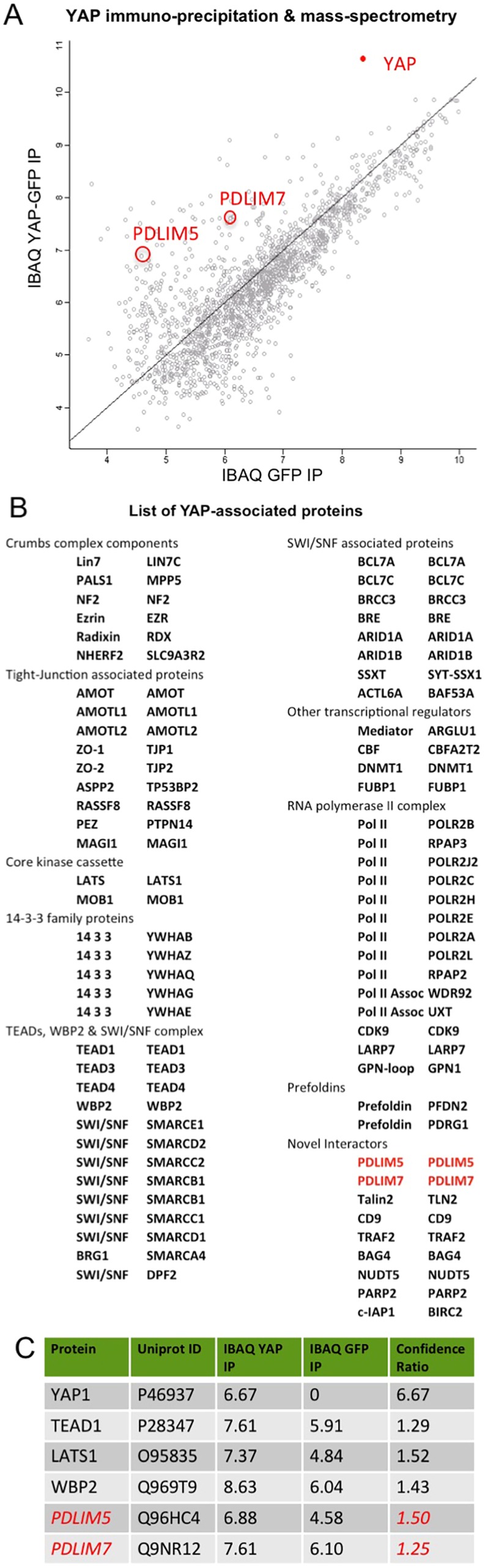
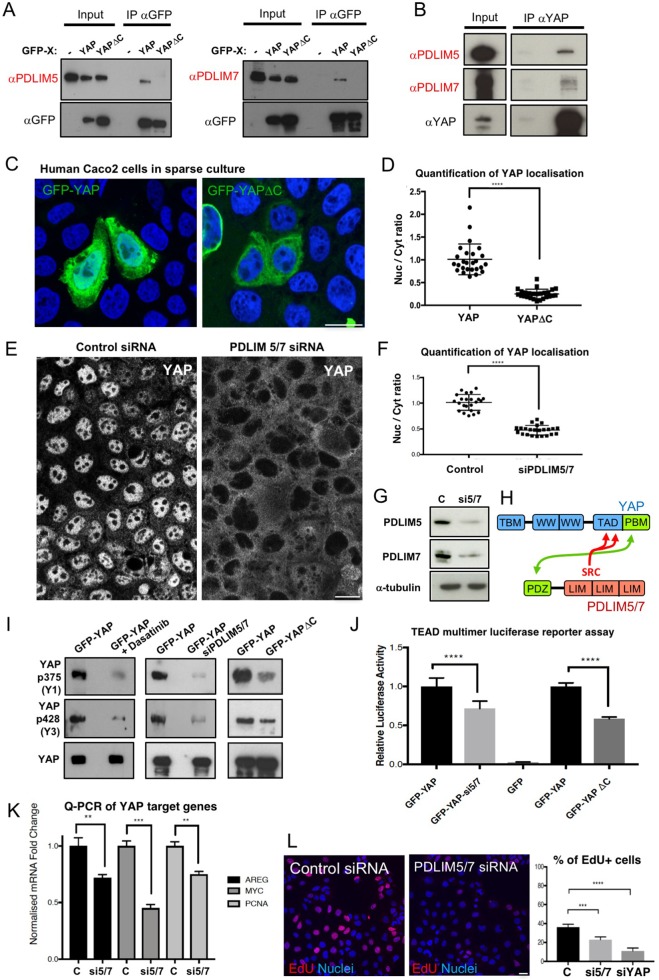
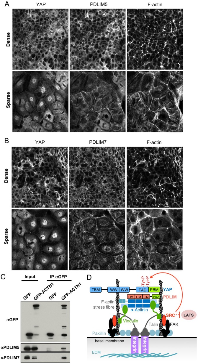
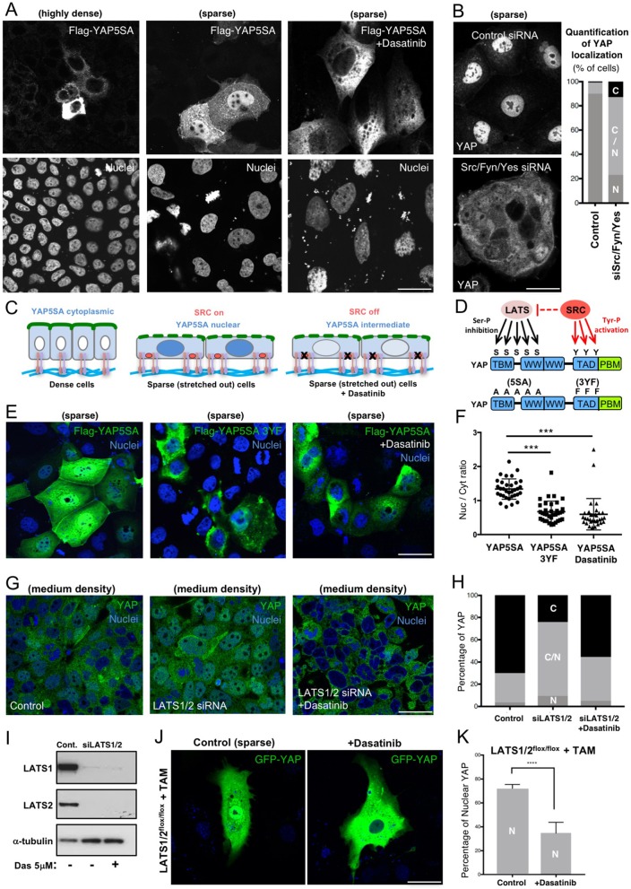
Human cells can sense mechanical stress acting upon integrin adhesions and respond by sending the YAP (also known as YAP1) and TAZ (also known as WWTR1) transcriptional co-activators to the nucleus to drive TEAD-dependent transcription of target genes. How integrin signaling activates YAP remains unclear. Here, we show that integrin-mediated mechanotransduction requires the Enigma and Enigma-like proteins (PDLIM7 and PDLIM5, respectively; denoted for the family of PDZ and LIM domain-containing proteins). YAP binds to PDLIM5 and PDLIM7 (hereafter PDLIM5/7) via its C-terminal PDZ-binding motif (PBM), which is essential for full nuclear localization and activity of YAP. Accordingly, silencing of PDLIM5/7 expression reduces YAP nuclear localization, tyrosine phosphorylation and transcriptional activity. The PDLIM5/7 proteins are recruited from the cytoplasm to integrin adhesions and F-actin stress fibers in response to force by binding directly to the key stress fiber component α-actinin. Thus, forces acting on integrins recruit Enigma family proteins to trigger YAP activation during mechanotransduction.This article has an associated First Person interview with the first author of the paper.

Keywords: Enigma; Hippo; Integrin; Mechanotransduction; PDLIM5; PDLIM7; YAP.


Conflict of interest statement

Competing interestsThe authors declare no competing or financial interests.

Figures

Fig. 1.
Fig. 1.
YAP immunoprecipitation and mass-spectrometry analysis of binding partners. (A) YAP IP-MS analysis of co-precipitated proteins identifies the Engima family proteins PDLIM5 and PDLIM7 as novel YAP-associated proteins. Axes are log10-transformed values. (B) List of all YAP-associated proteins identified in the experiment shown in A. (C) Comparison of confidence ratios for PDLIM5/7 and known YAP interactors.
Fig. 2.
Fig. 2.
The Enigma family proteins PDLIM5/7 bind to the YAP C-terminal PBM and promote YAP nuclear localization and transcriptional activity. (A) Confirmation of YAP–PDLIM5 and YAP–PDLIM7 interaction by co-immunoprecipitation (IP) of GFP-tagged YAP, and immunoblotting with anti-PDLIM5 and anti-PDLIM7 antibodies. Both Enigma family proteins PDLIM5 and PDLIM7 bind to the YAP C-terminal PBM, as deletion of this motif (YAPΔC) abolishes the interaction in co-immunoprecipitation experiments. (B) Endogenous YAP co-immunoprecipitates with PDLIM5 and PDLIM7. (C) Deletion of the C-terminal PBM (YAPΔC) reduces nuclear localization of GFP-tagged YAP in human Caco2 cells plated at low density. Results in A–C are representative of n=3 biological replicates. (D) Quantification (mean±s.d., n=3) of YAP localization for experiments as shown in C. ****P<0.001 (Student's t-test). (E) Double-silencing of both PDLIM5 and PDLIM7 expression in human Caco2 cells strongly reduces nuclear localization of YAP. Results are representative of n=6 biological replicates. (F) Quantification (mean±s.e.m.) of YAP localization for experiments as shown in E. ****P<0.001 (Student's t-test). (G) Confirmation of depletion of PDLIM5 and PDLIM7 expression levels for the siRNA (si5/7) treatment used in E. C, control siRNA. Results are representative of n=3 biological replicates. (H) Schematic diagram of the interaction between the YAP PBM and the PDZ domain of PDLIM5/7 proteins. Proximity of Src phosphorylation sites to the PBM is shown. (I) Silencing of PDLIM5/7 in human Caco2 cells reduces tyrosine phosphorylation of YAP. Results are representative of n=3 biological replicates. (J) Silencing of PDLIM5/7 in human Caco2 cells reduces YAP-driven TEAD transcriptional activity, as measured by a TEAD multimer luciferase reporter assay (relative to a Renilla luciferase control) (mean±s.e.m., n=10 from two independent experiments). Similar results were obtained upon deletion of the YAP C-terminal PBM. ****P<0.001 (Student's t-test). (K) Silencing of PDLIM5/7 in human Caco2 cells reduces expression of the YAP-target genes AREG, MYC and PCNA (mean±s.e.m., n=3). **P<0.01, ***P<0.005 (Student's t-test). (L) Silencing of PDLIM5/7 in human Caco2 cells reduces the rate of cell proliferation, as measured through a pulse of EdU incorporation. Upon loss of PDLIM5/7, proliferation slows by approximately half compared with control cells, which is comparable in magnitude to that achieved by silencing of YAP itself (siYAP). Results in the graph are mean±s.d. (n=3). ****P<0.001, ***P<0.005 (Student's t-test). Scale bars: 20 µm (C,E,L).
Fig. 3.
Fig. 3.
PDLIM5/7 proteins localize in the cytoplasm in dense cells but to basal stress fibers in sparse cells and bind directly to the stress fiber component α-actinin 1. (A) PDLIM5 localizes to the cytoplasm in dense cells but translocates, in part, to F-actin stress fibers and adherens junctions in sparsely plated cells. (B) PDLIM7 localizes to the cytoplasm in dense cells but translocates in part to F-actin stress fibers and adherens junctions in sparsely plated cells. (C) Co-immunoprecipitation of PDLIM5/7 proteins with GFP-tagged α-actinin 1 from human Caco2 cells. Results are representative of n=3 biological replicates. (D) Schematic diagram of YAP recruitment via the Enigma PDLIM5/7 proteins to integrin–Src signaling complexes to sense mechanical forces basally. Scale bar: 20 µm (A,B).
Fig. 4.
Fig. 4.
Mechanical control of YAP can occur independently of the canonical LATS phosphorylation on serine residues seen in the Hippo pathway and involves Src family kinase phosphorylation on tyrosine residues. (A) Mutation of five serine residues in YAP (5SA), to render it not capable of being phosphorylated by LATS kinase, does not lead to constitutively nuclear localization in human Caco2 cells. Flag-tagged YAP5SA responds to changes in cell density, becoming cytoplasmic in highly dense cultures and nuclear in sparse cultures with spread-out cells. The nuclear localization of YAP5SA is dependent on Src family kinases, as it is reduced upon treatment with the Src inhibitor Dasatinib. Results are representative of n=4 biological replicates. (B) Silencing of Src/Fyn/Yes kinases by triple siRNA causes a reduction in YAP nuclear localization. A quantification of the percentage of cells with each localization category (N, nuclear; N/C, nuclear and cytoplasmic; C, cytoplasmic) is shown on the right, n=3. (C) Schematic diagram of YAP5SA subcellular localization (blue) in response to cell density and upon treatment with Dasatinib in (A). Basal integrin attachments to the ECM are shown. (D) Schematic diagram of YAP phosphorylation by LATS1/2 and Src family kinases, and of serine (S) to alanine (A), and tyrosine (Y) to phenylalanine (F) mutations. (E) Flag-tagged YAP5SA nuclear localization depends upon three tyrosine residues in its transcriptional activation domain (TAD), whose mutation to phenylalanine (3YF) reduces nuclear localization in a manner similar to that seen upon treatment with Dasatinib. Results are representative of n=3 biological replicates. (F) Quantification of results from the experiment in E (mean±s.e.m., n=3). (G) YAP immunostaining (green) of human Caco2 epithelial cells at medium density transfected with either scrambled control siRNAs or LATS1/2 siRNAs (siLATS1/2) in the presence or absence of the Src family kinase inhibitor Dasatinib. DAPI marks nuclei (blue). Results are representative of n=3 biological replicates. (H) Quantification of results from the experiment in G (n=3). (I) Confirmation that LATS1/2 siRNAs, as used in experiment in G, effectively reduce LATS1 and LATS2 protein levels. (J) LATS1/2 double floxed MEFs transfected with Cre-ERt and YAP before treatment with Tamoxifen and Dasatinib to induce deletion of both LATS1 and LATS2 genes. (K) Quantification of YAP nuclear localization in experiments as in J (mean±s.e.m.; n=2 biological replicates each counting at least 600 cells from 8–10 independent areas over many coverslips). ****P<0.001 (Student's t-test). Scale bars: 20 μm (B), 30 μm (A,E), 50 μm (G,J).

References

    1. Avizienyte E. and Frame M. C. (2005). Src and FAK signalling controls adhesion fate and the epithelial-to-mesenchymal transition. Curr. Opin. Cell Biol. 17, 542-547. 10.1016/j.ceb.2005.08.007 - DOI - PubMed
    1. Badouel C., Gardano L., Amin N., Garg A., Rosenfeld R., Le Bihan T. and McNeill H. (2009). The FERM-domain protein Expanded regulates Hippo pathway activity via direct interactions with the transcriptional activator Yorkie. Dev. Cell 16, 411-420. 10.1016/j.devcel.2009.01.010 - DOI - PubMed
    1. Benham-Pyle B. W., Pruitt B. L. and Nelson W. J. (2015). Cell adhesion. Mechanical strain induces E-cadherin-dependent Yap1 and beta-catenin activation to drive cell cycle entry. Science 348, 1024-1027. 10.1126/science.aaa4559 - DOI - PMC - PubMed
    1. Chen Z., Oh D., Dubey A. K., Yao M., Yang B., Groves J. T. and Sheetz M. (2018). EGFR family and Src family kinase interactions: mechanics matters? Curr. Opin. Cell Biol. 51, 97-102. 10.1016/j.ceb.2017.12.003 - DOI - PubMed
    1. Dupont S., Morsut L., Aragona M., Enzo E., Giulitti S., Cordenonsi M., Zanconato F., Le Digabel J., Forcato M., Bicciato S. et al. (2011). Role of YAP/TAZ in mechanotransduction. Nature 474, 179-183. 10.1038/nature10137 - DOI - PubMed

Publication types