. 2018 Dec 10;131(24):jcs222034.
doi: 10.1242/jcs.222034.

An interaction network between the SNARE VAMP7 and Rab GTPases within a ciliary membrane-targeting complex

Affiliations

An interaction network between the SNARE VAMP7 and Rab GTPases within a ciliary membrane-targeting complex

Vasundhara Kandachar et al. J Cell Sci. .

Abstract

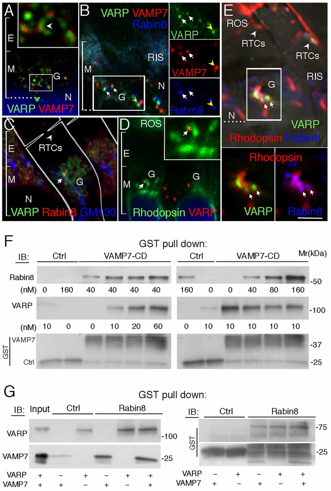
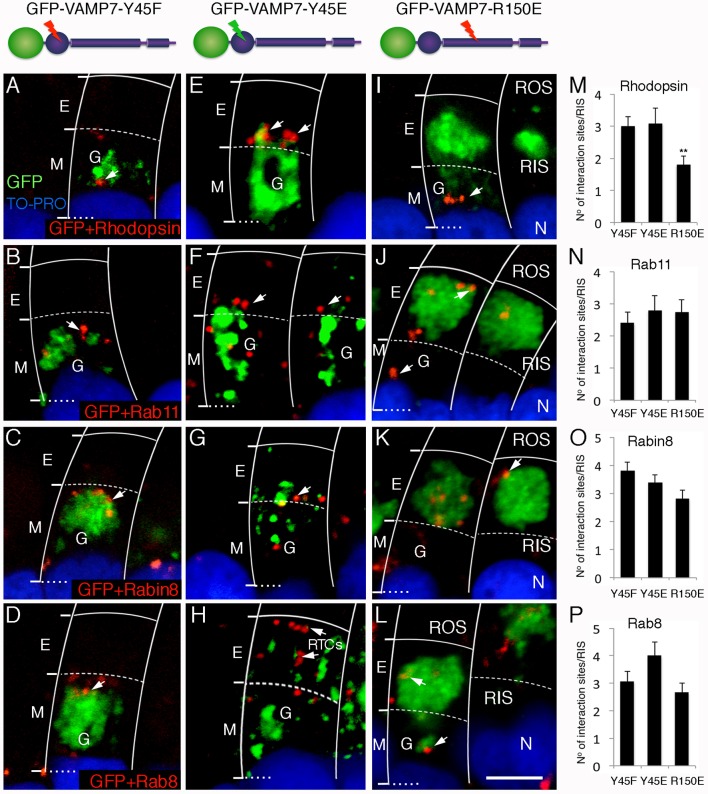
The Arf4-rhodopsin complex (mediated by the VxPx motif in rhodopsin) initiates expansion of vertebrate rod photoreceptor cilia-derived light-sensing organelles through stepwise assembly of a conserved trafficking network. Here, we examine its role in the sorting of VAMP7 (also known as TI-VAMP) - an R-SNARE possessing a regulatory longin domain (LD) - into rhodopsin transport carriers (RTCs). During RTC formation and trafficking, VAMP7 colocalizes with the ciliary cargo rhodopsin and interacts with the Rab11-Rabin8-Rab8 trafficking module. Rab11 and Rab8 bind the VAMP7 LD, whereas Rabin8 (also known as RAB3IP) interacts with the SNARE domain. The Arf/Rab11 effector FIP3 (also known as RAB11FIP3) regulates VAMP7 access to Rab11. At the ciliary base, VAMP7 forms a complex with the cognate SNAREs syntaxin 3 and SNAP-25. When expressed in transgenic animals, a GFP-VAMP7ΔLD fusion protein and a Y45E phosphomimetic mutant colocalize with endogenous VAMP7. The GFP-VAMP7-R150E mutant displays considerable localization defects that imply an important role of the R-SNARE motif in intracellular trafficking, rather than cognate SNARE pairing. Our study defines the link between VAMP7 and the ciliary targeting nexus that is conserved across diverse cell types, and contributes to general understanding of how functional Arf and Rab networks assemble SNAREs in membrane trafficking.

Keywords: Cilium; Rab GTPase; Rhodopsin; SNARE; Sensory receptor; VAMP7.


Conflict of interest statement

Competing interestsThe authors declare no competing or financial interests.

Figures

Fig. 1.
Fig. 1.
VAMP7 is localized at the Golgi/TGN and on ciliary-targeted RTCs, and forms a complex with syntaxin 3 and SNAP-25. (A) Diagram of the photoreceptor cell. Ciliary targeting of rhodopsin through the RIS proceeds from the Golgi (G) in the myoid region (M), through the ellipsoid region (E), which is crowded with mitochondria, to the base of the cilium (arrow), for the delivery to ROS. AJ, adherens junction. (B) Confocal optical section of a retina labeled with mouse (m) anti-VAMP7 (green), annotated as in A. G, Golgi; m, mitochondria. The dotted line represents the outer limiting membrane (OLM), comprising adherens junctions (AJ) between photoreceptors and retinal glial cells (Müller cells). Nuclei (N) were stained with TO-PRO-3 (blue). (C) Two confocal sections from a Z-stack of a retina labeled with anti-VAMP7 (m) (green) and rabbit (r) anti-ASAP1 (red). Scale bar: 5 µm (for B), 7 µm (for C). (D) Following pulse-chase labeling and retinal subcellular fractionation, the distribution of rhodopsin was determined by SDS-PAGE and autoradiography (of [35S]rhodopsin). A duplicate gel was tested by immunoblotting on a single blot, as indicated. (E–G) VAMP7+rhodopsin interaction sites (red dots) detected by PLA using anti-rhodopsin mAb11D5(m) and anti-VAMP7(m) conjugated to PLUS and MINUS probes, respectively, as described in the Materials and Methods (E). PLA for VAMP7(m)+ASAP1(r) (F). PLA for VAMP7(m) and phosphorylated tyrosine (p-tyr)(r) (G). (H–J) PLA with a single antibody to rhodopsin (H), ASAP1 (I) or VAMP7 (J). No red fluorescence was detected. (K,L) PLA for VAMP7(m) and syntaxin 3 (STX3)(r) (K), or SNAP-25(r) (L). Cells were visualized by DIC. Arrows in K and L indicate sites of colocalization. Scale bar: 5 µm (for E–G,K,L), 10 µm (H–J). (M) Post-nuclear supernatant (PNS) (0.1 retina) was immunoprecipitated (IP) with anti-syntaxin 3(r) and IPs were analyzed by immunoblotting (IB) as indicated.
Fig. 2.
Fig. 2.
In the presence of rhodopsin, VAMP7 associates with components of the Rab11–Rabin8–Rab8 ciliogenesis cascade. The R150E mutation in VAMP7 does not prevent its interaction with the components of the Rab11–Rabin8–Rab8 ciliogenesis cascade. (A–F) PLA in control (A,C,E) and cycloheximide-treated (B,D,F) retinas: VAMP7(m)+Rab11(r) (A,B), VAMP7(m)+Rabin8(r) (C,D) and VAMP7(m)+Rab8(r) (E,F). (G) For each PLA pair, interaction sites were counted (five transgene-expressing photoreceptors were counted from 10 sections from different Z-stacks; 50 cells) and the data were expressed as a means±s.e.m. (n=50) percentage of the total interaction sites within the RIS. ***P<1.6×10⁻¹⁸ (Student's t-test). (H) Retinas labeled with antibody to rhodopsin (m) and Rab6 (r). Arrow highlights the presence vs absence of rhodopsin in the Golgi. Scale bars: 5 µm. (I) A GST fusion protein containing the cytosolic domain of VAMP7 (V7-CD) or GST alone (Ctrl), was incubated with 6His–Rab8, in the presence of GDPβS or GTPγS, with or without 6His–Rabin8. Bound Rabin8 and Rab8 were detected by immunoblotting (IB). The GST fusion proteins were detected with anti-GST antibody. (J) V7-CD or Ctrl, was incubated with or without 6His–Rabin8, GTPγS-bound Rab11a or the C-terminal end of FIP3 (FIP3F1). Bound proteins were detected as above. (K) V7-CD and its mutants Y45E, R150, Y45E/R150E, ΔLD, ΔLD/R150E and LD, were incubated either with purified recombinant proteins, or with post-nuclear supernatant (PNS), and bound proteins were detected as indicated. GST fusion proteins were detected by anti-GST antibody (see Fig. S1). (L) GST–Rabin8 N (aa 1–235) and -Rabin8 C (aa 236–460) were incubated with purified VAMP7. Bound proteins were detected as above. (M) A model of the putative Rab11–Rabin8–VAMP7 complex generated by Dr Esben Lorentzen (Department of Molecular Biology and Genetics, Aarhus University, Aarhus, Denmark) with PyMOL, using the structure of the SRP receptor (PDB code 2FH5) (Schlenker et al., 2006), Rab11–Rabin8–FIP3 complex (PDB code 4UJ3) (Vetter et al., 2015a) and the VAMP7–VARP complex (PDB code: 4B93) (Schäfer et al., 2012). Positions of FIP3 and VARP in the crystalized complexes are indicated in light gray. VAMP7 SNARE domain, LD, and residues Y45 and R150 are indicated. M, myoid region; E, ellipsoid region; G, Golgi, N, nucleus; m, mouse antibody; r rabbit antibody.
Fig. 3.
Fig. 3.
Rabin8 cooperates with VARP in ciliary trafficking of VAMP7. (A–E) Retinas labeled with anti-VARP (r) and anti-VAMP7 (m) (A); anti-VAMP7 (m) (red), anti-Rabin8 (r) (blue), followed by anti-VARP conjugated to Alexa Fluor 488 (green) (B); anti-Rabin8 (r) (red) and anti-GM130(m) (blue) and anti-VARP-488 (green) (C); anti-rhodopsin 11D5 (m) (green) and anti-VARP (r) (red) (D); anti-rhodopsin 11D5 (m) (red), anti-Rabin8(r) (blue) and anti-VARP-488 (green) (E). The boxed area is repeated in insets to show individual staining patterns separately. Arrows in B indicate carriers containing VAMP7, whereas a yellow arrowhead indicates carries that appear in the process of fusion; arrow in C indicates the Golgi; arrows in D indicate nascent RTCs. Scale bar: 5 µm for A–E and 2.5 µm for insets in A and D. (F) GST–VAMP7 CD, or GST alone (Ctrl), was incubated with 6His–Rabin8 followed by increasing concentrations of purified recombinant VARP or vice versa. Bound proteins were immunoblotted (IB), as indicated. (G) GST–Rabin8 or GST alone (Ctrl) was incubated with or without recombinant VARP and/or VAMP7. Bound proteins and GST fusion proteins were detected as above. M, myoid region; E, ellipsoid region; G, Golgi, N, nucleus; m, mouse antibody; r rabbit antibody.
Fig. 4.
Fig. 4.
GFP–VAMP7 is present in the Golgi and on RTCs in transgenic Xenopus laevis photoreceptors. (A–C) Transgenic eyes expressing GFP–VAMP7 (A), GFP–VAMP7-LD (B) and GFP–VAMP7-ΔLD (C) (green). Glycosylated membrane proteins are detected by WGA (red). Nuclei are stained with DAPI (blue). (D–I) Transgenic retinas stained with WGA. Different transgene expression levels are indicated by: + (low), ++ (medium) and +++ (high). Arrowheads indicate RIS plasma membrane. (J,L,N) Transgenic retinas were labeled with anti-rhodopsin mAb 11D5 (red). Asterisk in J indicates site of GFP–VAMP7 accumulation in the Golgi, whereas arrows indicate tips of the trans-Golgi cisternae. (K,M,O) PLA between rhodopsin (m) and anti-GFP (r) detecting GFP–VAMP7 (K), GFP–VAMP7-LD (M) and GFP–VAMP7-ΔLD (O). Arrows in K indicate sites of interaction. Retinal sections were visualized by DIC in K, M and O. Scale bar: 500 µm (for A–C), 15 µm (for D–J and N), 10 µm (for K–M,O). G, Golgi; m, mouse antibody; r, rabbit antibody.
Fig. 5.
Fig. 5.
Distribution of GFP–VAMP7 mutants in transgenic photoreceptors. (A–E) Transgenic expression of GFP–VAMP7-Y45F (A), -Y45E (B), -R150E (C), -Y45E/R150E (D) and -ΔLD/R150E (E) in retinal photoreceptors (green). Sections are stained with WGA and DAPI, as in Fig. 4. CP, calycal processes, the villous structures that originate from the RIS plasma membrane and surround the base of the ROS. (F–J) GFP-VAMP7 mutants (green) and WGA-positive membranes (red), partitioning into distinct membranes. White arrows indicate WGA–GFP colocalization, whereas green arrows indicate GFP-positive WGA-negative membranes. RIS plasma membrane domains are indicated by white arrowheads (juxtaciliary) and yellow arrowheads (lateral). Scale bar: 10 µm (for A–E); 7 µm (for F–J). G, Golgi, N, nucleus.
Fig. 6.
Fig. 6.
Distinct interaction sites of GFP–VAMP7 mutants with rhodopsin, and components of the Rab11–Rabin8–Rab8 ciliogenesis cascade. (A–D) Interaction sites (red dots) identified by PLA between anti-GFP (r) detecting GFP–VAMP7-Y45F and anti-rhodopsin mAb11D5 (m) (A), or anti-Rab11 (m) (B); anti-GFP (m) and anti-Rabin8 (r) (C) or anti-Rab8 (r) (D). (E–H) PLA between anti-GFP (r) detecting GFP-VAMP7-Y45E and anti-rhodopsin mAb11D5(m) (E), or anti-Rab11(m) (F); anti-GFP(m) and anti-Rabin8(r) (G) or anti-Rab8(r) (H). (I–L) PLA between anti-GFP (r) detecting GFP–VAMP7-R150E and anti-rhodopsin mAb11D5(m) (I), or anti-Rab11 (m) (J); anti-GFP (m) and anti-Rabin8 (r) (K) or anti-Rab8 (r) (L). Scale bar: 5 µm. Gamma adjustment was applied in panels E–I in order to reveal fainter GFP features that were not apparent in the uncorrected images. Arrows indicate sites of interaction. (M–P) Transgenic animals were analyzed at 2 months of age and 10-–20 confocal optical sections were generated for each PLA pair. Representative confocal sections encompassing the entire RIS were selected and interaction sites were counted in 15 photoreceptors for each PLA pair, analyzed using Student's t-test (n=15) and presented as the means±s.e.m. **P=0.006, Y45F versus R150E. M, myoid region; E, ellipsoid region; G, Golgi, N, nucleus; m, mouse antibody; r, rabbit antibody.
Fig. 7.
Fig. 7.
Aberrant vesicular structures containing GFP–VAMP7-R150E are deficient in typical regulators of intracellular trafficking. (A–C) Transgenic X. laevis expressing the GFP–VAMP7-R150E mutant and examined by CLEM. Retinas were first examined by confocal microscopy (A) and then processed and analyzed by EM (B). Six cells in the confocal optical section (a–f) were matched to the cells (a–f) in the EM micrograph. The asterisk marks a control photoreceptor (f), not expressing GFP–VAMP7-R150E. The boxed area in B is magnified in C. m, mitochondria, Ly, lysosome. (D–F) Transgenic retinas expressing GFP–VAMP7-R150E fusion protein (green) labeled with anti-LAMP1 (r) (D), anti-GFP (r) (E), or anti-βCOP (r) (F). Arrows point to localization of the proteins examined. (G) PLA between: Rab6 (r) and anti-GFP (m) detecting GFP–VAMP7-R150E. (H,I) EM of the photoreceptor cell expressing the R150E mutant (H). The boxed area is magnified in I. Arrows point to vesicular structures with electron-dense content. (J–M) Transgenic GFP–VAMP7-R150E retinas labeled with anti-IRBP (m) (J), anti-ASAP1 (r) (K), anti-FIP3 (r) (L) and anti-VARP (r) (M). (N) PLA between syntaxin 3 (r) and anti-GFP (m). (O) Retinas labeled with anti-peripherin (m) (red). (P,Q) The f1 generation of GFP–VAMP7-R150E-expressing retinas stained with WGA (red). (R) Labeling with anti-LC3 (r) (red, arrows). (S) Epithelial cells outside of Xenopus eye labeled with anti-LC3 (r) (red, arrows point to autophagosomes). (T,U) Labeling with anti-ATG16L1 (m) (red, arrow) (T), or anti-ubiquitin (m) (U). Cells were visualized by DIC. (V) The f1 generation of GFP-VAMP7-Y45E-expressing retinas stained with WGA (red). Scale bar: 25 µm (for A); 5 µm (for B,D–H); 10 µm (for J–N); 1 µm (for C,I); 12 µm (for O,R,S,T,U); 20 µm (for Q); 50 µm (for P,V). M, myoid region; E, ellipsoid region; G, Golgi, N, nucleus; m, mouse antibody; r, rabbit antibody.

Similar articles

Cited by

References

    1. Arshavsky V. Y. and Burns M. E. (2012). Photoreceptor signaling: supporting vision across a wide range of light intensities. J. Biol. Chem. 287, 1620-1626. 10.1074/jbc.R111.305243 - DOI - PMC - PubMed
    1. Bachmann-Gagescu R., Phelps I. G., Stearns G., Link B. A., Brockerhoff S. E., Moens C. B. and Doherty D. (2011). The ciliopathy gene cc2d2a controls zebrafish photoreceptor outer segment development through a role in Rab8-dependent vesicle trafficking. Hum. Mol. Genet. 20, 4041-4055. 10.1093/hmg/ddr332 - DOI - PMC - PubMed
    1. Bennett M. K., Garcia-Arrarás J. E., Elferink L. A., Peterson K., Fleming A. M., Hazuka C. D. and Scheller R. H. (1993). The syntaxin family of vesicular transport receptors. Cell 74, 863-873. 10.1016/0092-8674(93)90466-4 - DOI - PubMed
    1. Brown M. T., Andrade J., Radhakrishna H., Donaldson J. G., Cooper J. A. and Randazzo P. A. (1998). ASAP1, a phospholipid-dependent arf GTPase-activating protein that associates with and is phosphorylated by Src. Mol. Cell. Biol. 18, 7038-7051. 10.1128/MCB.18.12.7038 - DOI - PMC - PubMed
    1. Burgo A., Sotirakis E., Simmler M.-C., Verraes A., Chamot C., Simpson J. C., Lanzetti L., Proux-Gillardeaux V. and Galli T. (2009). Role of Varp, a Rab21 exchange factor and TI-VAMP/VAMP7 partner, in neurite growth. EMBO Rep. 10, 1117-1124. 10.1038/embor.2009.186 - DOI - PMC - PubMed

Publication types

MeSH terms