. 2018 Nov 7;285(1890):20181792.
doi: 10.1098/rspb.2018.1792.

Dental ontogeny in extinct synapsids reveals a complex evolutionary history of the mammalian tooth attachment system

Affiliations

Dental ontogeny in extinct synapsids reveals a complex evolutionary history of the mammalian tooth attachment system

Aaron R H LeBlanc et al. Proc Biol Sci. .

Abstract

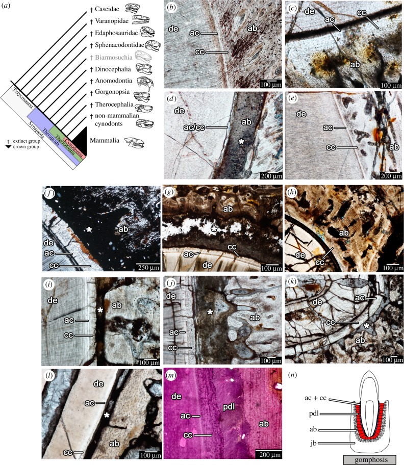
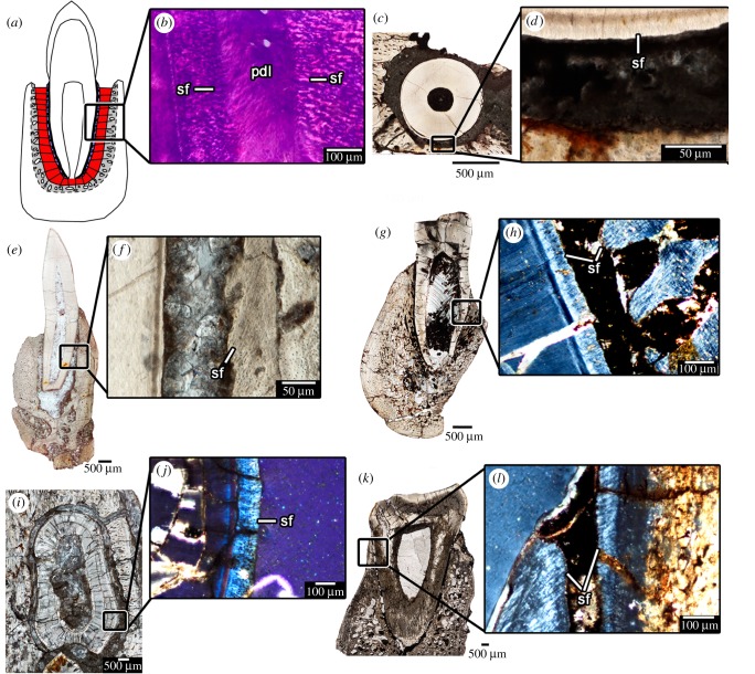
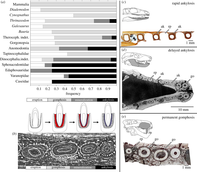
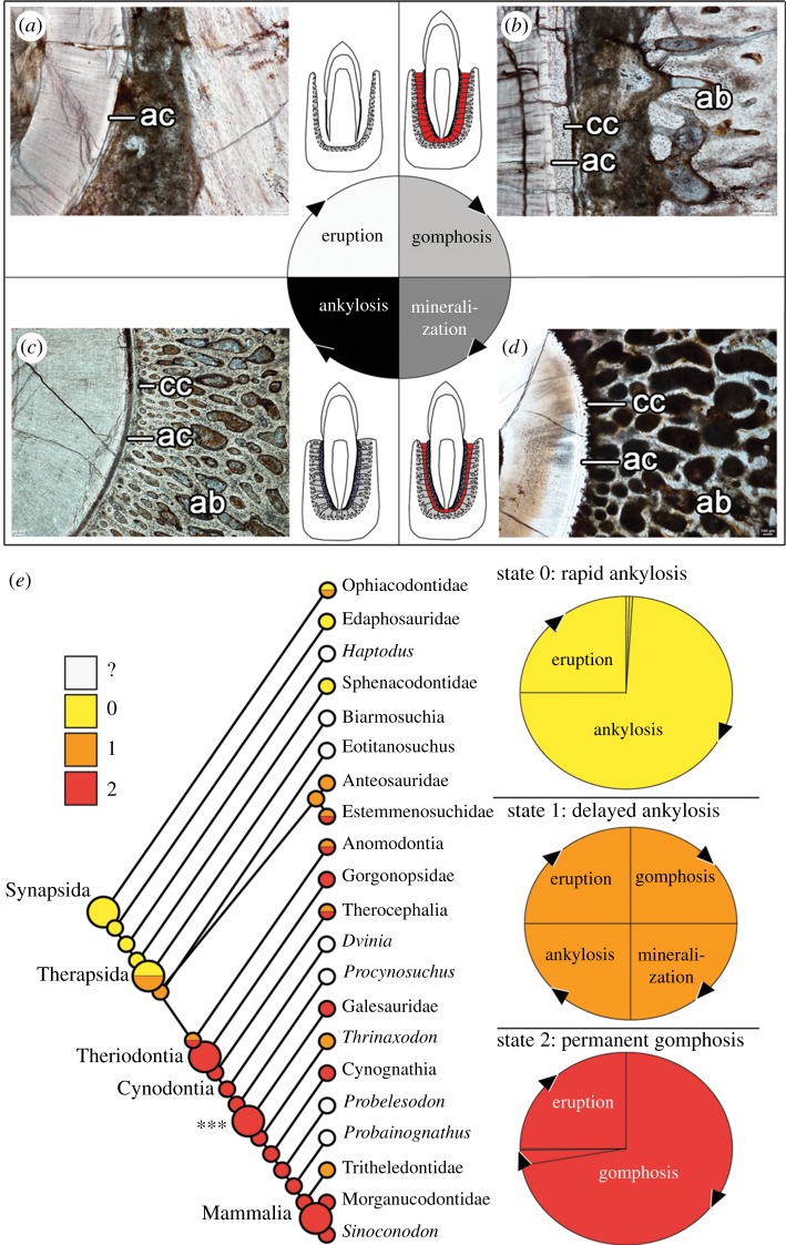
The mammalian dentition is uniquely characterized by a combination of precise occlusion, permanent adult teeth and a unique tooth attachment system. Unlike the ankylosed teeth in most reptiles, mammal teeth are supported by a ligamentous tissue that suspends each tooth in its socket, providing flexible and compliant tooth attachment that prolongs the life of each tooth and maintains occlusal relationships. Here we investigate dental ontogeny through histological examination of a wide range of extinct synapsid lineages to assess whether the ligamentous tooth attachment system is unique to mammals and to determine how it evolved. This study shows for the first time that the ligamentous tooth attachment system is not unique to crown mammals within Synapsida, having arisen in several non-mammalian therapsid clades as a result of neoteny and progenesis in dental ontogeny. Mammalian tooth attachment is here re-interpreted as a paedomorphic condition relative to the ancestral synapsid form of tooth attachment.

Keywords: ankylosis; dental histology; paedomorphosis; pelycosaur; therapsid.


Conflict of interest statement

The authors have no competing interests to declare.

Figures

Figure 1.
Figure 1.
Tooth attachment in stem and crown mammals (Synapsida). (a) Cladogram of the evolutionary relationships of the major taxa within Synapsida (modified from [5,20,25]). (b–l) thin sections showing tooth attachment tissues in major synapsid groups. (b) The sphenacodontid Dimetrodon (ROM 6039). (c) An indeterminate dinocephalian (BP/1/4851). (d) The anomodont Diictodon (ROM 52624). (e) A second individual of Diictodon (ROM 52624). (f) An indeterminate gorgonopsian (NMT RB404). (g) An indeterminate therocephalian (BP/1/7257). (h) The same therocephalian, but an adjacent tooth position. (i) The derived therocephalian Bauria (BP/1/2523). (j) The cynodont Cynognathus (BP/1/6097). (k) The cynodont Diademodon (BP/1/4652). (l) The extinct ungulate mammal Hyopsodus (USNM 595273). (m) A stained section of the periodontium in an extant badger (Taxidea). (n) Illustrated the arrangement of the attachment tissues of the gomphosis in Synapsida. ab, alveolar bone (grey); ac, acellular cementum (blue); cc, cellular cementum (blue); de, dentine; jb, jawbone; pdl, periodontal ligament (red). Asterisks indicate spaces formerly occupied by periodontal ligament in life.
Figure 2.
Figure 2.
‘Mammal’-like tooth attachment in non-mammalian therapsids. (a) Illustration of the tooth attachment tissues in a gomphosis. (b) Close-up of the tooth attachment tissues in an extant badger under cross-polarized light, showing the Sharpey's fibres of the PDL in the cementum and alveolar bone. (c) Transverse section of a tooth of a gorgonopsian (BP/1/784). (d) Close-up of the periodontal tissues in (c). (e) Coronal section of a tooth of an indeterminate therocephalian (BP/1/172). (f) Close-up of periodontal tissues in (e). (g) Coronal section of a tooth in the therocephalian Bauria (BP/1/2523). (h) Close-up of periodontal tissues in (g) under cross-polarized light. (i) Transverse section of a tooth in the cynodont Cynognathus (BP/1/6097). (j) Close-up of the periodontal tissues in (i) under cross-polarized light. (k) Coronal section of a tooth in the cynodont Diademodon (BP/1/4652). (l) Close-up of the periodontal tissues in (k) under cross-polarized light. pdl, periodontal ligament; sf, Sharpey's fibres.
Figure 3.
Figure 3.
Tooth attachment and relative frequency of tooth attachment stages. (a) The frequency of teeth at the eruption, gomphosis, mineralization and ankylosis stages in thin sections and CT scans of fossil synapsids, reflecting the proportion of time teeth spend in the respective stages (note: the mammal samples used here all had erupted permanent dentitions). (b) Digital transverse section through four postcanines of Thrinaxodon (BP/1/5372) showing successive stages of eruption, gomphosis, mineralization and ankylosis (from left to right). (c) varanopid dentary transverse section showing teeth at either the eruption stage or completely ankylosed (rapid ankylosis) (ROM 66866). (d) CT image of the lower jaws of the cynodont Thrinaxodon (BP/1/7199) showing teeth at eruption, gomphosis and ankylosis stages (delayed ankylosis). (e) gorgonopsian maxilla (BP/1/2395a) transverse section showing teeth either at the eruption or gomphosis stages (permanent gomphosis). ak, ankylosis; go, gomphosis, rp, resorption pit/erupting tooth. (Online version in colour.)
Figure 4.
Figure 4.
Heterochrony and the evolution of synapsid tooth attachment. (a) Eruption stage after the previous tooth is shed involves remodelling of tissues and formation of the tooth crown and early root tissues (image of erupting tooth of the cynodont Cynognathus) (BP/1/6097). (b) Gomphosis stage occurs when all three periodontal tissues are fully formed in a functional tooth (image of an erupted tooth root of Cynognathus) (BP/1/6097). (c) Mineralization stage occurs as alveolar bone begins to calcify centripetally. Minor centrifugal mineralization of cementum may also occur (image of a dinocephalian tooth root) (BP/1/6854). (d) Ankylosis stage occurs when alveolar bone and cementum meet and completely entomb the periodontal ligament (image of the tooth root of Dimetrodon) (ROM 6039). (e) Ancestral character state reconstruction for the major synapsid clades using a three-state character and the phylogeny of Sidor & Hopson [5]. Larger circles indicate the nodes for Synapsida, Therapsida, Theriodonta and Mammalia. Asterisks and associated large circle indicate earliest cynodont node for which histological data was actually observed. Small circles at terminal branches indicate character state codings for each OTU (operational taxonomic unit). ab, alveolar bone; ac, acellular cementum; cc, cellular cementum.

Similar articles

Cited by

References

    1. Crompton AW, Jenkins FA. 1968. Minor occlusion in Late Triassic mammals. Biol. Rev. 43, 427–458. (10.1111/j.1469-185X.1968.tb00966.x) - DOI - PubMed
    1. Weller JM. 1968. Evolution of mammalian teeth. J. Paleontol. 42, 268–290.
    1. Kemp TS. 1982. Mammal-like reptiles and the origin of mammals. London, UK: Academic Press Inc.
    1. Kemp TS. 2006. The origin and early radiation of the therapsid mammal-like reptiles: a palaeobiological hypothesis. J. Evol. Biol. 19, 1231–1247. (10.1111/j.1420-9101.2005.01076.x) - DOI - PubMed
    1. Sidor CA, Hopson JA. 1998. Ghost lineages and ‘mammalness’: assessing the temporal pattern of character acquisition in the Synapsida. Paleobiol. 24, 254–273.

Publication types