

Structure-guided development of selective M3 muscarinic acetylcholine receptor antagonists
- PMID: 30404914
- PMCID: PMC6255194
- DOI: 10.1073/pnas.1813988115
Structure-guided development of selective M3 muscarinic acetylcholine receptor antagonists
Abstract
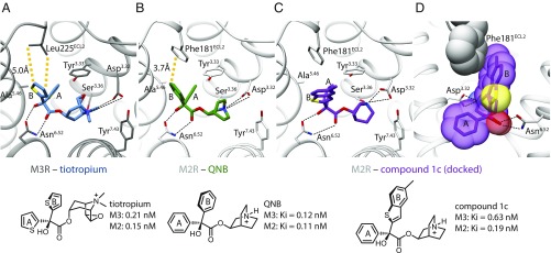
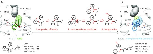
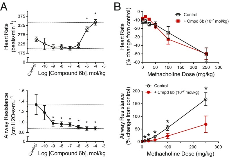
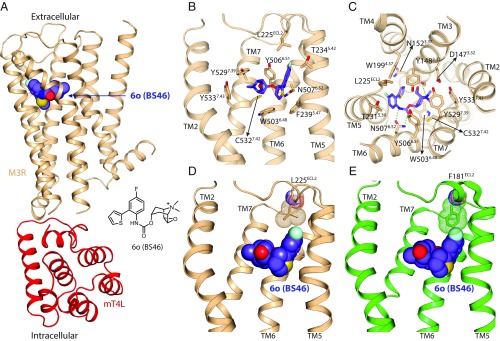
Drugs that treat chronic obstructive pulmonary disease by antagonizing the M3 muscarinic acetylcholine receptor (M3R) have had a significant effect on health, but can suffer from their lack of selectivity against the M2R subtype, which modulates heart rate. Beginning with the crystal structures of M2R and M3R, we exploited a single amino acid difference in their orthosteric binding pockets using molecular docking and structure-based design. The resulting M3R antagonists had up to 100-fold selectivity over M2R in affinity and over 1,000-fold selectivity in vivo. The crystal structure of the M3R-selective antagonist in complex with M3R corresponded closely to the docking-predicted geometry, providing a template for further optimization.
Keywords: G protein-coupled receptor; crystal structure; drug design; muscarinic receptor; subtype selectivity.
Copyright © 2018 the Author(s). Published by PNAS.
Conflict of interest statement
Conflict of interest statement: B.K.K. is a cofounder of and consultant for ConfometRx, Inc.
Figures
Similar articles
-
Novel Potent Muscarinic Receptor Antagonists: Investigation on the Nature of Lipophilic Substituents in the 5- and/or 6-Positions of the 1,4-Dioxane Nucleus.J Med Chem. 2020 Jun 11;63(11):5763-5782. doi: 10.1021/acs.jmedchem.9b02100. Epub 2020 May 19. J Med Chem. 2020. PMID: 32374602 Free PMC article.
-
Dibenzodiazepinone-type muscarinic receptor antagonists conjugated to basic peptides: Impact of the linker moiety and unnatural amino acids on M2R selectivity.Eur J Med Chem. 2021 Mar 5;213:113159. doi: 10.1016/j.ejmech.2021.113159. Epub 2021 Jan 13. Eur J Med Chem. 2021. PMID: 33571911
-
Muscarinic M₃ receptors contribute to allergen-induced airway remodeling in mice.Am J Respir Cell Mol Biol. 2014 Apr;50(4):690-8. doi: 10.1165/rcmb.2013-0220OC. Am J Respir Cell Mol Biol. 2014. PMID: 24156289
-
Novel insights into M3 muscarinic acetylcholine receptor physiology and structure.J Mol Neurosci. 2014 Jul;53(3):316-23. doi: 10.1007/s12031-013-0127-0. Epub 2013 Sep 26. J Mol Neurosci. 2014. PMID: 24068573 Free PMC article. Review.
-
Muscarinic acetylcholine receptor X-ray structures: potential implications for drug development.Curr Opin Pharmacol. 2014 Jun;16:24-30. doi: 10.1016/j.coph.2014.02.006. Epub 2014 Mar 21. Curr Opin Pharmacol. 2014. PMID: 24662799 Free PMC article. Review.
Cited by
-
Daidzein is the in vivo active compound of Puerariae Lobatae Radix water extract for muscarinic receptor-3 inhibition against overactive bladder.Front Pharmacol. 2022 Oct 4;13:924251. doi: 10.3389/fphar.2022.924251. eCollection 2022. Front Pharmacol. 2022. PMID: 36267287 Free PMC article.
-
Deepening insights into cholinergic agents for intraocular pressure reduction: systems genetics, molecular modeling, and in vivo perspectives.Front Mol Biosci. 2024 Jul 26;11:1423351. doi: 10.3389/fmolb.2024.1423351. eCollection 2024. Front Mol Biosci. 2024. PMID: 39130374 Free PMC article.
-
Heterogeneous Dielectric Implicit Membrane Model for the Calculation of MMPBSA Binding Free Energies.J Chem Inf Model. 2019 Jun 24;59(6):3041-3056. doi: 10.1021/acs.jcim.9b00363. Epub 2019 Jun 13. J Chem Inf Model. 2019. PMID: 31145610 Free PMC article.
-
Novel Potent Muscarinic Receptor Antagonists: Investigation on the Nature of Lipophilic Substituents in the 5- and/or 6-Positions of the 1,4-Dioxane Nucleus.J Med Chem. 2020 Jun 11;63(11):5763-5782. doi: 10.1021/acs.jmedchem.9b02100. Epub 2020 May 19. J Med Chem. 2020. PMID: 32374602 Free PMC article.
-
Xanomeline displays concomitant orthosteric and allosteric binding modes at the M4 mAChR.Nat Commun. 2023 Sep 6;14(1):5440. doi: 10.1038/s41467-023-41199-5. Nat Commun. 2023. PMID: 37673901 Free PMC article.
References
-
- Hulme EC, Birdsall NJM, Buckley NJ. Muscarinic receptor subtypes. Annu Rev Pharmacol Toxicol. 1990;30:633–673. - PubMed
-
- Wess J. Molecular biology of muscarinic acetylcholine receptors. Crit Rev Neurobiol. 1996;10:69–99. - PubMed
-
- Caulfield MP, Birdsall NJM. International Union of Pharmacology. XVII. Classification of muscarinic acetylcholine receptors. Pharmacol Rev. 1998;50:279–290. - PubMed
Publication types
MeSH terms
Substances
Associated data
- Actions
Grants and funding
LinkOut - more resources
Full Text Sources
Other Literature Sources
