. 2018 Nov 7;9(1):4670.
doi: 10.1038/s41467-018-07036-w.

Dynamic control of proinflammatory cytokines Il-1β and Tnf-α by macrophages in zebrafish spinal cord regeneration

Affiliations

Dynamic control of proinflammatory cytokines Il-1β and Tnf-α by macrophages in zebrafish spinal cord regeneration

Themistoklis M Tsarouchas et al. Nat Commun. .

Abstract

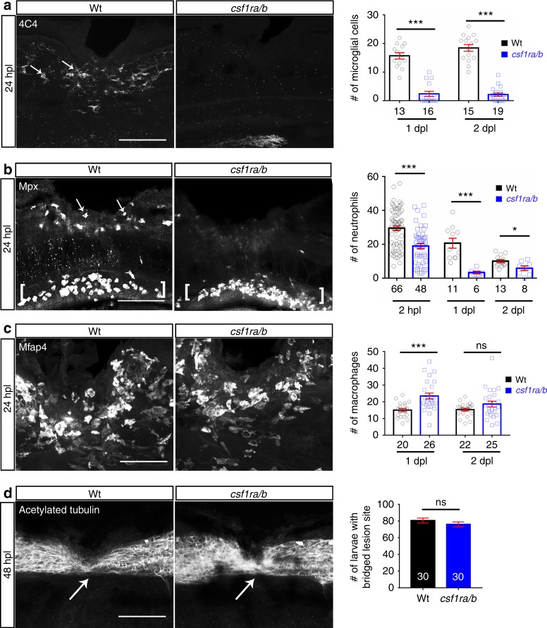
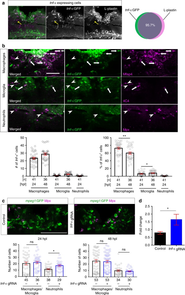
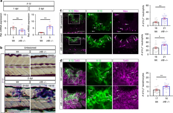
Spinal cord injury leads to a massive response of innate immune cells in non-regenerating mammals, but also in successfully regenerating zebrafish. However, the role of the immune response in successful regeneration is poorly defined. Here we show that inhibiting inflammation reduces and promoting it accelerates axonal regeneration in spinal-lesioned zebrafish larvae. Mutant analyses show that peripheral macrophages, but not neutrophils or microglia, are necessary for repair. Macrophage-less irf8 mutants show prolonged inflammation with elevated levels of Tnf-α and Il-1β. Inhibiting Tnf-α does not rescue axonal growth in irf8 mutants, but impairs it in wildtype animals, indicating a pro-regenerative role of Tnf-α. In contrast, decreasing Il-1β levels or number of Il-1β+ neutrophils rescue functional regeneration in irf8 mutants. However, during early regeneration, interference with Il-1β function impairs regeneration in irf8 and wildtype animals. Hence, inflammation is dynamically controlled by macrophages to promote functional spinal cord regeneration in zebrafish.


Conflict of interest statement

The authors declare no competing interests.

Figures

Fig. 1
Fig. 1
Spinal injury leads to an inflammatory response that promotes axonal regeneration. a Neutrophils, macrophages, and microglial cells show different dynamics after injury. Neutrophils (Mpx+) accumulate in the injury site very early, peaking at 2 hpl. Macrophages (mpeg1:GFP+/4C4) and microglial cell (mpeg1:GFP+/4C4+) numbers peak at 48 hpl. Fluorescence images were projected onto transmitted light images. be Incubation with dexamethasone (timeline in b) reduces neutrophil and macrophage numbers (c, d; Mann–Whitney U-test: **P < 0.01, ***P < 0.001), as well as the proportion of animals with axonal bridging (e; Fisher’s exact test: ***P < 0.001). fi, Incubation of animals with LPS during early regeneration (timeline in f) increased numbers of neutrophils and macrophages (g, h; t-test: **P < 0.01, ***P < 0.001), as well as the proportion of animals with axonal bridging at 24 hpl (i Fisher’s exact test: *P < 0.05). Lateral views of the injury site are shown; rostral is left. Rectangles indicate region of quantification; arrows indicate axonal bridging. Scale bars: 50 μm; Error bars indicate SEM
Fig. 2
Fig. 2
In the irf8 mutant, axonal regeneration and functional recovery after injury show long-term impairment. a In situ hybridisation for mpeg1 confirms the absence of macrophages and microglial cells before and after injury in the irf8 mutant compared to controls. Arrows indicate labelling around the injury site and brackets indicate the ventral area of the larvae where the macrophages can be found in the circulation. Note that blackish colour is due to melanocytes. b Quantification of the proportion of larvae with axonal bridging (anti-acetylated Tubulin) shows that at 1 dpl, axonal bridging is unimpaired in irf8 mutants, whereas at 2 dpl, irf8 mutants fail to show full regrowth and even by 5 dpl, the proportion of irf8 larvae with a bridged lesion site is still lower than in wildtype controls (Fisher’s exact test: ***P < 0.001, n.s. indicated no significance). c Irf8 mutants never fully recover touch-evoked swimming distance in the observation period, whereas wildtype control animals do. Representative swim tracks are displayed. Note that unlesioned irf8 larvae show swimming distances that are comparable to those in wildtype controls (Two-way ANOVA: F15,1372 = 11.42, P < 0.001; unles. = unlesioned, les. = lesioned). All lesions are done at 3 dpf. Lateral views of the injury site are shown; rostral is left. Arrows indicate axonal bridging. Scale bars: 200 μm in a and 50 μm in c. Error bars indicate SEM
Fig. 3
Fig. 3
Absence of microglial cells and reduced neutrophil numbers do not affect axon bridging. a Numbers of microglial cells (4C4+; arrows) in the injury site of the csf1ra/b mutants are much lower than in wildtype animals (t-test: ***P < 0.001). b Fewer neutrophils (Mpx+) are found in the injury site (arrows) of csf1ra/b mutants than in wildtype animals (t-test: *P < 0.05, ***P < 0.001). Note neutrophils ventral to the injury site (brackets). c The number of macrophages (Mfap4+) is increased in the injury site in the mutants at 1 dpl, but not at 2 dpl (t-test: ***P < 0.001, ns indicates no significance). d Immunostaining against acetylated tubulin shows that axon bridging (arrows) is not affected in the mutants compared to wildtype animals at 2 dpl (Fisher’s exact test: ns indicates no significance). Lateral views of the injury site are shown; rostral is left. Wt = wildtype; Scale bars: 50 µm in a, b, d; 25 µm in b. Error bars indicate SEM
Fig. 4
Fig. 4
Inflammation is bi-phasic and dysregulated in irf8 mutants. a, b Absence of macrophages in the irf8 mutant fish leads to increased il- and tnf-α mRNA levels during the late stage of inflammation (>12 hpl). An early peak in tnf-α expression is missing in irf8 mutants. c, d Expression of anti-inflammatory cytokines, tgf-β1a and tgf-β3, which peak during late regenerative phases in wildtype animals, is strongly reduced in irf8 mutants (t-tests: *P < 0.05, **P < 0.01, ***P < 0.001; wt = wildtype animals). # indicates statistical significance when compared to unlesioned animals. Error bars indicate SEM
Fig. 5
Fig. 5
Tnf-α is essential for axonal regeneration. a Tnf-α inhibition by Pomalidomide reduces the proportion of wildtype animals with axon bridging at 1 and 2 dpl. No effect is observed in irf8 mutants (Two-way ANOVA followed by Bonferroni post-test: F3,16 = 12.16, **P < 0.01, n.s indicates no significance). b CRISPR/Cas9-mediated disruption of tnf-α is effective as shown by RFLP analysis. This reveals efficient somatic mutation in the gRNA target site, indicated by resistance to restriction endonuclease digestion (arrow). c Axonal bridging (arrow; Xla.Tubb:DsRed+) is strongly impaired after disruption of the tnf-α gene. (Fisher’s exact test: ***P < 0.001) and the impairment persists at 5 dpl. Lateral views of the injury site are shown; rostral is left. Scale bar: 50 μm. Error bars indicate SEM
Fig. 6
Fig. 6
Tnf-α is expressed by macrophages and regulates the immune response. a Top row: tnf-α:GFP labelling occurs almost exclusively in L-plastin+ immune cells (L-plastin in green; tnf-α:GFP in magenta; yellow arrow indicates a rare tnf- α:GFP+ microglial cell; 12 hpl) b In the injury site, the number and proportion of macrophages (Mfap4+) that are tnf-α:GFP+ are much higher than numbers and proportions of microglia (4C4+) and neutrophils (Mpx+), indicating that the main source of Tnf-α is the macrophages. Arrows indicate double-labelled cells and arrowheads indicate immune cells that are tnf-α:GFP-. Single optical sections are shown; the proportion of macrophages that are tnf-α:GFP+ decreases over time, whereas the proportion of tnf-α:GFP+ microglial cells slightly increases (One-way ANOVA followed by Bonferroni post-test: F4,195 = 376.3, **P < 0.01, *P < 0.05). c Quantification of the immune cells after tnf-α gRNA injection shows that Tnf-α disruption leads to increased numbers of neutrophils (Mpx+) at 1 dpl but not at 2 dpl, whereas the numbers of macrophages/microglia (mpeg1:GFP+) remains unchanged (Mann–Whitney U-test: *P < 0.05, ns indicates no significance). d qRT-PCR indicates that tnf-α disruption leads to increased levels of il- mRNA at 2 dpl (t-tests: *P < 0.05). Lateral views of the injury site are shown; rostral is left. Scale bars: 50 μm. Error bars indicate SEM
Fig. 7
Fig. 7
Inhibition of Il-1β function rescues axonal regeneration in irf8 mutants. Lateral views of the injury site are shown; rostral is left. a YVAD reduces expression levels of il- and tnf-α in irf8 mutants (two-sample t-test: *P < 0.05) at 2 dpl. b YVAD impairs migration of peripheral macrophages (Mfap4+) and neutrophils (Mpx+) in wildtype animals and irf8 mutants (only neutrophils quantified, due to absence of macrophages) (t-tests: ***P < 0.001). c YVAD moderately reduces the number of TUNEL+ cells in the irf8 mutants at 2 dpl. (Two-Way ANOVA followed by Bonferroni multiple comparisons: F3,121 = 112.5, ***P < 0.001). d YVAD does not influence axonal regeneration in wildtype animals but rescues axonal bridging (arrows) in irf8 mutants (Fisher’s exact test: **P < 0.01, ns indicates no significance) at 2 dpl. e Impaired touch-evoked swimming distance in irf8 mutants is rescued by YVAD treatment, to levels that are no longer different from lesioned and unlesioned wildtype animals at 2 dpl. YVAD has no influence on swimming distance in lesioned or unlesioned wildtype animals (Two-way ANOVA followed by Bonferroni multiple comparisons: F1,309 = 35.229, ***P < 0.0001, ns indicates no significance). Rectangle in b denotes quantification area. Scale bar: 50 μm for b, d. Error bars indicate SEM
Fig. 8
Fig. 8
Levels of il- expression are increased in the injury site of irf8 mutants. a At 1 dpl, expression levels of il- are comparable between irf8 mutants and wildtype (Wt) animals but are higher in the mutant at 2 dpl in qRT-PCR (t-test: **P < 0.01, ns indicates no significance). b In situ hybridisation confirms increased expression of il- mRNA at 2 dpl. c In the injury site, the number and proportion of neutrophils (Mpx+) that are Il-1β immuno-positive (arrows) are increased in irf8 mutants at 1 dpl compared to wildtype animals. d The number of basal keratinocytes (Tp63+) that are Il-1β immuno-positive is increased in irf8 mutants. Single optical sections are shown; boxed areas are shown in higher magnifications (t-test: *P < 0.05, **P < 0.01). Lateral views of the injury site are shown; rostral is left. Scale bars: 100 μm in b, c, d and 50 µm for higher magnification areas. Error bars indicate SEM
Fig. 9
Fig. 9
Preventing neutrophil formation partially rescues functional spinal cord regeneration in the irf8 mutant. a In irf8 mutants, higher peak numbers of neutrophils (Mpx+) at 2 hpl and slower clearance over the course of regeneration are observed (Two-Way ANOVA followed by Bonferroni multiple comparisons: F8,427 = 13.19 *P < 0.05, **P < 0.01, ***P < 0.001). Note that wildtype data are the same as shown in Fig. 1a, as counts in irf8 mutants and wildtype animals were done in the same experiments. b Combination treatment with pu.1 and gcsfr morpholinos efficiently prevents neutrophil accumulation in the lesion site (Mann–Whitney U-test: ***P < 0.001). c In pu.1/gcsfr morpholino injected irf8 mutant fish, levels of il- and tnf-α mRNA expression are reduced at 2 dpl, as shown by qRT-PCR (t-test: ***P < 0.001). d, e In pu.1/gcsfr morpholino injected irf8 mutant fish, axonal bridging (arrows, d Fisher’s exact test: **P < 0.01) and behavioural recovery (e One-Way ANOVA followed by Bonferroni multiple comparisons: F5,142 = 23.21, **P < 0.01, ns indicates no significance) are partially rescued. Lateral views of the injury site are shown; rostral is left. Scale bars: 100 μm. Error bars indicate SEM

References

    1. Wehner D, et al. Wnt signaling controls pro-regenerative Collagen XII in functional spinal cord regeneration in zebrafish. Nat. Commun. 2017;8:126. doi: 10.1038/s41467-017-00143-0. - DOI - PMC - PubMed
    1. Becker T, Wullimann MF, Becker CG, Bernhardt RR, Schachner M. Axonal regrowth after spinal cord transection in adult zebrafish. J. Comp. Neurol. 1997;377:577–595. doi: 10.1002/(SICI)1096-9861(19970127)377:4<577::AID-CNE8>3.0.CO;2-#. - DOI - PubMed
    1. Kigerl KA, et al. Identification of two distinct macrophage subsets with divergent effects causing either neurotoxicity or regeneration in the injured mouse spinal cord. J. Neurosci. 2009;29:13435–13444. doi: 10.1523/JNEUROSCI.3257-09.2009. - DOI - PMC - PubMed
    1. Norden DM, Muccigrosso MM, Godbout JP. Microglial priming and enhanced reactivity to secondary insult in aging, and traumatic CNS injury, and neurodegenerative disease. Neuropharmacology. 2015;96:29–41. doi: 10.1016/j.neuropharm.2014.10.028. - DOI - PMC - PubMed
    1. Taoka Y, et al. Role of neutrophils in spinal cord injury in the rat. Neuroscience. 1997;79:1177–1182. doi: 10.1016/S0306-4522(97)00011-0. - DOI - PubMed

Publication types