. 2018 Nov 7;8(1):16478.
doi: 10.1038/s41598-018-34605-2.

Dynamic recognition and linkage specificity in K63 di-ubiquitin and TAB2 NZF domain complex

Affiliations

Dynamic recognition and linkage specificity in K63 di-ubiquitin and TAB2 NZF domain complex

Kei Moritsugu et al. Sci Rep. .

Abstract

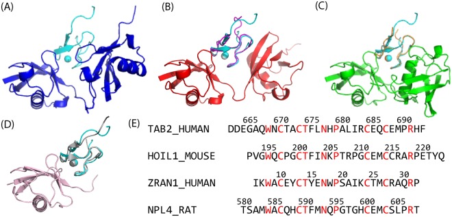
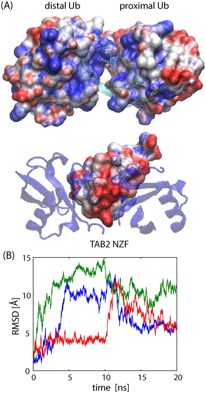
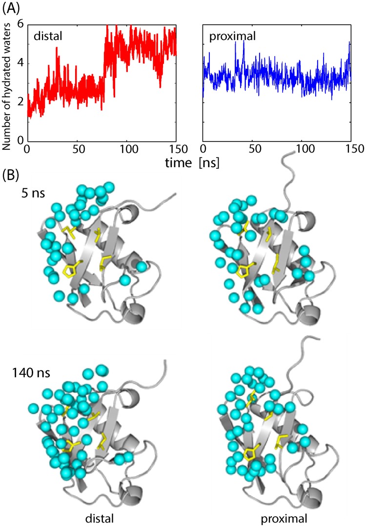
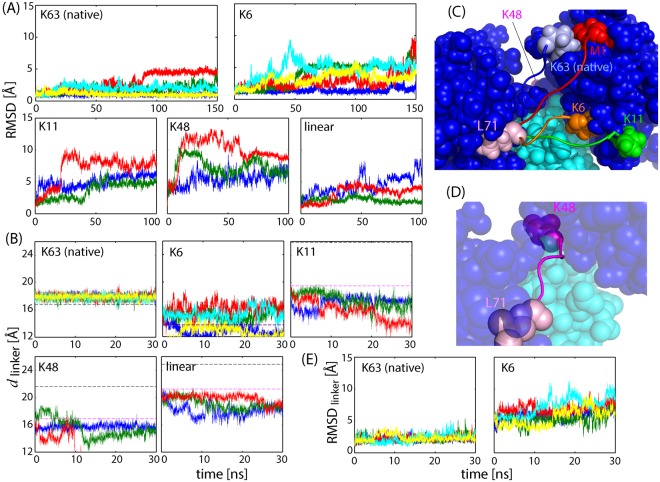
Poly-ubiquitin (poly-Ub) is involved in various cellular processes through the linkage-specific recognition of Ub-binding domains (UBD). In this study, using molecular dynamics (MD) simulation together with an enhanced sampling method, we demonstrated that K63-linked di-Ub recognizes the NZF domain of TAB2, a zinc finger UBD, in an ensemble of highly dynamic structures that form from the weak interactions between UBD and the flexible linker connecting the two Ubs. However, the K63 di-Ub/TAB2 NZF complex showed a much more compact and stable ensemble than the non-native complexes, linear di-Ub/TAB2 NZF and K33 di-Ub/TAB2 NZF, that were modeled from linear di-Ub/HOIL-1L NZF and K33 di-Ub/TRABID NZF1, respectively. We further demonstrated the importance of the length and position of the Ub-Ub linker in the results of MD simulations of K63 di-Ub/TAB2 NZF by changing the Ub linkage from the native K63 to four different non-native linkages, linear, K6, K11, and K48, while maintaining inter-molecular contacts in the native complex. No systems with non-native linkage maintained the native binding configuration. These simulation results provide an atomistic picture of the linkage specific recognition of poly-Ubs leading to the biological functions such as cellular colocalization of various component proteins in the signal transduction pathways.


Conflict of interest statement

The authors declare no competing interests.

Figures

Figure 1
Figure 1
Crystal structures of the di-Ub/NZF complexes and sequence alignment of the NZF domains. (A) K63 di-Ub/TAB2 NZF (PDB: 2wwz). (B) Linear di-Ub/HOIL-1L NZF Δtail (PDB: 3b0a). Here, Δtail indicates that the NZF domain does not contain the C-terminal helix of HOIL-1L (224–249). (C) K33 di-Ub/TRABID NZF1 (PDB: 5af6). (D) Mono Ub/NPL4 NZF (PDB: 1q5w). In (BD), TAB2 NZF (cyan) is drawn after superimposition to the core region (1–70) of the distal Ub (left moiety of di-Ub) or mono-Ub. (E) The sequences of the NZF domains used in this study were aligned. ZRAN1 corresponds to TRABID. Red characters indicate the amino acids which are conserved in all four proteins.
Figure 2
Figure 2
FES of K63 di-Ub/TAB2 NZF. (A) FES plotted on a two-dimensional space, Cα RMSD values of TAB2 NZF, and di-Ub from the crystal structure (PDB: 2wwz) after superimposing Cα atoms of the core regions of di-Ub (1–70). The inset is the same plot but calculated from five 150-ns MD simulations (Fig. S1A). The lower free energy region (<10 KJ/mol) is named as the “near-native” ensemble, while the higher free energy region (>10 KJ/mol) is named as the “non-native” ensemble. (B) The FES for the subset with the condition that TAB2 NZF contacts (any non-hydrogen atom pairs whose distance is less than 4 Å) both distal and proximal Ubs. (C) The same as (A) but plotted on the two Cα RMSD values of TAB2 NZF, one superimposing the distal Ub (1–70) and the other superimposing the proximal Ub (1–70). (D) Three representative structures of the complex whose position in the FES are indicated by the numbers in (A,C).
Figure 3
Figure 3
Electrostatic interactions between K63 di-UB and TAB2 NZF. (A) Surfaces of the electrostatic potential of K63 di-Ub without TAB2 NZF and isolated TAB2 NZF, drawn by VMD. (B) Time courses of Cα RMSD for three MD simulations (colored in red, blue, and green) of free K63 di-Ub (core regions, 1–70) starting from the crystal structure of the complex after removing TAB2 NZF.
Figure 4
Figure 4
Hydration of the interface between di-Ub and TAB2 NZF. (A) Number of hydrated water molecules found on the interface (waters within 4 Å of the protein interface). The left and right figures are for the distal Ub and proximal Ub, respectively. (B) The snapshots of interfacial waters for the distal Ub (left) and proximal Ub (right) after 5 ns (upper) and 140 ns (lower) during MD simulation. The water molecules are depicted by cyan spheres and the four residues of the I44 patch (L8, I44, H68, and V70) are drawn as yellow sticks.
Figure 5
Figure 5
MD simulations of mono-Ub/TAB2 NZF. (A) Time courses of Cα RMSD values for three 150-ns MD simulations (colored in red, blue, and green) from the crystal structure of distal Ub/TAB2 NZF (PDB: 2wwz as the reference structure). The blue curve overshoots the upper range of the figure. (B) Those of proximal Ub/TAB2 NZF. (C) Those of mono-Ub/Npl4 NZF (PDB: 1q5w).
Figure 6
Figure 6
MD simulations of linear di-Ub/TAB2 NZF and K33 di-Ub/TAB2 NZF. (A) FES of linear di-Ub/TAB2 NZF obtained in the MSES simulation, plotted on a two-dimensional space, Cα RMSD values of TAB2 NZF and di-Ub from the initial homology model, after superimposing Cα atoms of di-Ub (1–70). (B) The same as (A), but plotted on the two distances, dR42-Q686 and dE140-R683. (C) Two representative structures whose positions in the FES are indicated in (A,B). TAB2 NZF changes the position by rotation while maintaining the salt bridge, R72-E688. (D) Time courses of Cα RMSD value of K33 di-Ub/TAB2 NZF from the initial homology model, after superimposing Cα atoms of di-Ub (1–70), obtained in the three 150-ns MD simulations (colored in red, blue, and green). The blue curve overshoots the range of the figure. (E) Two representative structures indicated in (D).
Figure 7
Figure 7
MD simulations of native K63-diUb/TAB2 NZF complex and artificially linked K6, K11, K48 di-UB/TAB2 NZF complexes. (A) Cα RMSD during the simulations of the five di-Ub/TAB2 NZF complexes (native K63: 5 × 150 ns; K6: 5 × 150 ns; K11, K48 and linear: 3 × 100 ns) from each initial model after superimposing Cα atoms of di-Ub (1–70). Note that the initial model for K63 is the crystal structure (PDB: 2wwz), but that the other models are different from each other depending on the modeling procedures (see Methods for details). (B) The linker length, dlinker, or distance between Cα of L71 and Nζ of each Lys residues (either K63, K6, K11, or K48 di-Ub) or N of M1 for linear di-Ub. The first 30 ns data are shown here. The horizontal black and pink lines are the dlinker values for the crystal structure and for the initial models, respectively; these values are different from each other depending on the linkage. (C) The initial models for MD simulations: di-Ub: blue; TAB2 NZF: cyan; linkers (71–76) and lysine residues (or M1): light blue (K63), red (linear), orange (K6), and green (K11). The linker of K48 (pink) is not drawn here because it is located on the other side of the proximal Ub. (D) The initial model for the K48 complex. (E) Cα RMSD of the Ub-Ub linker (71–76) for K63 and K6 di-Ub/TAB2 NZF.

Similar articles

Cited by

References

    1. Husnjak K, Dikic I. Ubiquitin-binding proteins: Decoders of ubiquitin-mediated cellular functions. Annu. Rev. Biochem. 2012;81:291–322. doi: 10.1146/annurev-biochem-051810-094654. - DOI - PubMed
    1. Komander D, Rape M. The ubiquitin code. Annu. Rev. Biochem. 2012;81:203–229. doi: 10.1146/annurev-biochem-060310-170328. - DOI - PubMed
    1. Chen ZJ, Sun LJ. Nonproteolytic functions of ubiquitin in cell signaling. Mol. Cell. 2009;33:275–286. doi: 10.1016/j.molcel.2009.01.014. - DOI - PubMed
    1. Akutsu M, Dikic I, Bremm A. Ubiquitin chain diversity at a glance. J. Cell Sci. 2016;129:875–880. doi: 10.1242/jcs.183954. - DOI - PubMed
    1. Yau R, Rape M. The increasing complexity of the ubiquitin code. Nat. Cell Biol. 2016;18:579–586. doi: 10.1038/ncb3358. - DOI - PubMed

Publication types

MeSH terms