. 2018 Oct 24:12:766.
doi: 10.3389/fnins.2018.00766. eCollection 2018.

Axonal Injury Induces ATF3 in Specific Populations of Sacral Preganglionic Neurons in Male Rats

Affiliations

Axonal Injury Induces ATF3 in Specific Populations of Sacral Preganglionic Neurons in Male Rats

Agnes W Wong et al. Front Neurosci. .

Abstract

Compared to other neurons of the central nervous system, autonomic preganglionic neurons are unusual because most of their axon lies in the periphery. These axons are vulnerable to injury during surgical procedures, yet in comparison to peripheral neurons and somatic motor neurons, the impact of injury on preganglionic neurons is poorly understood. Here, we have investigated the impact of axotomy on sacral preganglionic neurons, a functionally diverse group of neurons required for micturition, defecation, and sexual function. We have previously observed that after axotomy, the injury-related transcription factor activating transcription factor-3 (ATF3) is upregulated in only half of these neurons (Peddie and Keast, 2011: PMID: 21283532). In the current study, we have investigated if this response is constrained to particular subclasses of preganglionic neurons that have specific functions or signaling properties. Seven days after unilateral pelvic nerve transection, we quantified sacral preganglionic neurons expressing ATF3, many but not all of which co-expressed c-Jun. This response was independent of soma size. Subclasses of sacral preganglionic neurons expressed combinations of somatostatin, calbindin, and neurokinin-1 receptor, each of which showed a similar response to injury. We also found that in contrast to thoracolumbar preganglionic neurons, the heat shock protein-25 (Hsp25) was not detected in naive sacral preganglionic neurons but was upregulated in many of these neurons after axotomy; the majority of these Hsp25 neurons expressed ATF3. Together, these studies reveal the molecular complexity of sacral preganglionic neurons and their responses to injury. The simultaneous upregulation of Hsp25 and ATF3 may indicate a distinct mechanism of regenerative capacity after injury.

Keywords: axon regeneration; axotomy; intermediolateral nucleus; parasympathetic; preganglionic neurons; spinal nerve injury.


Figures

FIGURE 1
FIGURE 1
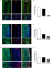
Expression of ATF3 and c-Jun in L6-S1 preganglionic neurons one week after unilateral transection of the pelvic nerve. Representative horizontal sections of spinal cord were immunolabeled for c-Jun (green) and ATF3 (red). Preganglionic neurons were identified by their uptake of retrograde tracer [FluoroGold (FG), here colorized blue]. Because all of the preganglionic neurons are quite intensely labeled by FluoroGold (shown as blue), the c-Jun nuclei appear turquoise rather than green and the ATF3 nuclei appear pink rather than red. Images are oriented with rostral at the top and lateral on the left (spared) or right (injured) of the relevant panel. (A) Contralateral to the injury (“spared”), no preganglionic neurons were immunoreactive for ATF3 and rare nuclei were immunoreactive for c-Jun; in contrast, ipsilateral to injury (“injured”) many preganglionic neurons were immunoreactive for ATF3, c-Jun, or both transcription factors (ATF3/c-Jun). An example of each is shown by an arrowhead (c-Jun), short arrow (ATF3), and long arrow (ATF3/c-Jun). (B) Higher magnification of spared (left) and injured (right) sides of the L6-S1 IML. (C) Quantitation of FG-positive preganglionic neurons ipsilateral to injury, showing the proportion that are immunoreactive for either ATF3 or c-Jun alone, or for both transcription factors (ATF3/c-Jun); together these comprise 56.1 ± 2.8% of sacral preganglionic neurons identified by FG (n = 6). (D) No difference was identified between the soma profile areas of neurons expressing ATF3 (153 ± 9 μm2) and ATF3-negative neurons (162 ± 20 μm2). Data shown as (D) mean soma profile area from each side of each animal and (E) individual neurons pooled from all animals. n = 4 rats, minimum of 20 neurons measured on each side in each rat, two-tailed, paired t-test: P = 0.65. Calibration bars represent 100 μm.
FIGURE 2
FIGURE 2
Expression of somatostatin (SOM), calbindin (CAL), and neurokinin-1 receptor (NK1R) in L6-S1 preganglionic neurons. Representative horizontal sections of spinal cord immunolabeled for pairs of neural markers are shown in (A–C); higher magnification images from different sections are provided in the corresponding lower panels. Preganglionic neurons were identified by ChAT-immunoreactivity, here colorized blue. Images are oriented with rostral at the top and lateral on the right. SOM-, CAL-, and NK1R-immunoreactivity were each identified in subpopulations of preganglionic neurons. Immunoreactivity for SOM and CAL was cytoplasmic, NK1R-immunoreactivity was primarily in the plasma membrane and CAL-immunoreactive preganglionic neurons showed cytoplasmic or nuclear labeling, or both. SOM- and NK1R-immunoreactive dendrites extended toward the midline, and also labeled many neuronal somata near the midline. CAL-immunoreactive somata, presumed interneurons, were found between the intermediolateral column and the midline; these consistently showed cytoplasmic labeling. (D–F) Analysis of expression patterns in preganglionic neurons. (D) Subpopulations of neurons expressed either SOM or CAL, but very few expressed both SOM and CAL (SOM/CAL) (n = 6 rats). (E) Subpopulations of neurons expressed either NK1R or CAL, with few expressing both NK1R and CAL (NK1R/CAL). In total, 45.0 ± 2.4% of sacral preganglionic neurons expressed NK1R and 24.9 ± 1.5% expressed CAL (n = 5 rats). (F) Subpopulations of neurons expressed either NK1R or SOM, with some neurons expressing both NK1R and SOM (NK1R/SOM). In total, 33.1 ± 1.3% of preganglionic neurons expressed NK1R and 33.0 ± 1.3% expressed SOM (n = 6 rats). Calibration bars represent 100 μm.
FIGURE 3
FIGURE 3
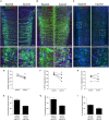
Expression of ATF3 in somatostatin (SOM), neurokinin-1 receptor (NK1R), and calbindin (CAL) subpopulations of L6-S1 preganglionic neurons one week after unilateral transection of the pelvic nerve. Representative horizontal sections of spinal cord immunolabeled for SOM, NK1R, or CAL (green) or ATF3 (red) are shown in A–C; higher magnification images from the same images are provided in the lower panels of each, as indicated by the boxes. Preganglionic neurons were identified by their uptake of retrograde tracer [FluoroGold (FG), here colorized blue]. Images are oriented with rostral at the top and lateral on the left (spared) or right (injured) of the relevant panel. Calibration bars represent 100 μm. ATF3 was only expressed in neurons on the injured side of the cord. (D) Injury reduced the proportion of preganglionic neurons expressing SOM (paired two-tailed t-test, P = 0.003; n = 5 rats). (E) Around half of the injured SOM-positive preganglionic neurons expressed ATF3; these comprised around one-third of the entire ATF3-positive population. (F) Injury reduced the proportion of preganglionic neurons expressing NK1R (paired two-tailed t-test, P = 0.043; n = 5 rats). (G) Around half of the injured NK1R-positive preganglionic neurons expressed ATF3; these comprised around one-third of the entire ATF3-positive population. (H) No effect of injury was detected on the proportion of preganglionic neurons expressing CAL (paired two-tailed t-test, P = 0.542; n = 5 rats). (I) Around half of the injured CAL-positive preganglionic neurons expressed ATF3; these comprised around one-third of the entire ATF3-positive population.
FIGURE 4
FIGURE 4
Expression of heat shock protein 25 (Hsp25) in lumbar and sacral preganglionic neurons. (A,B) Representative horizontal sections of spinal cord, oriented with rostral at the top and lateral on the right. (A) Preganglionic neurons in the intermediolateral column of the thoracolumbar spinal cord (T13-L2) were identified by immunoreactivity for choline acetyltransferase (ChAT, blue); a subpopulation of these neurons showed strong immunoreactivity for Hsp25 (green). (B) In the L6-S1 spinal cord, many preganglionic neurons (identified by FluoroGold labeling, colorized blue here) showed strong immunoreactivity for Hsp25 one week after axotomy; many of these Hsp25-immunoreactive neurons also expressed ATF3. In contrast, the uninjured (spared) side of the L6-S1 IML expressed neither Hsp25 nor ATF3. (C,D) Quantitation of L6-S1 preganglionic neurons after axotomy, showing that the majority of neurons upregulating Hsp25 also express ATF3, and that about one-third of ATF3 neurons showed upregulation of Hsp25 (n = 6). Calibration bars represent 100 μm.

Similar articles

Cited by

References

    1. Anderson C. R., Edwards S. L. (1994). Intraperitoneal injections of Fluorogold reliably labels all sympathetic preganglionic neurons in the rat. J. Neurosci. Methods 53 137–141. 10.1016/0165-0270(94)90170-8 - DOI - PubMed
    1. Antal M., Freund T. F., Polgár E. (1990). Calcium-binding proteins, parvalbumin- and calbindin-D 28k-immunoreactive neurons in the rat spinal cord and dorsal root ganglia: a light and electron microscopic study. J. Comp. Neurol. 295 467–484. 10.1002/cne.902950310 - DOI - PubMed
    1. Arms L., Vizzard M. A. (2011). Neuropeptides in lower urinary tract function. Handb. Exp. Pharmacol. 202 395–423. 10.1007/978-3-642-16499-6_19 - DOI - PMC - PubMed
    1. Averill S., Michael G. J., Shortland P. J., Leavesley R. C., King V. R., Bradbury E. J., et al. (2004). NGF and GDNF ameliorate the increase in ATF3 expression which occurs in dorsal root ganglion cells in response to peripheral nerve injury. Eur. J. Neurosci. 19 1437–1445. 10.1111/j.1460-9568.2004.03241.x - DOI - PubMed
    1. Barber R. P., Phelps P. E., Houser C. R., Crawford G. D., Salvaterra P. M., Vaughn J. E. (1984). The morphology and distribution of neurons containing choline acetyltransferase in the adult rat spinal cord: an immunocytochemical study. J. Comp. Neurol. 229 329–346. 10.1002/cne.902290305 - DOI - PubMed