. 2018 Oct 18:12:41.
doi: 10.3389/fnsys.2018.00041. eCollection 2018.

Functional Diversity of Thalamic Reticular Subnetworks

Affiliations

Functional Diversity of Thalamic Reticular Subnetworks

John W Crabtree. Front Syst Neurosci. .

Abstract

The activity of the GABAergic neurons of the thalamic reticular nucleus (TRN) has long been known to play important roles in modulating the flow of information through the thalamus and in generating changes in thalamic activity during transitions from wakefulness to sleep. Recently, technological advances have considerably expanded our understanding of the functional organization of TRN. These have identified an impressive array of functionally distinct subnetworks in TRN that participate in sensory, motor, and/or cognitive processes through their different functional connections with thalamic projection neurons. Accordingly, "first order" projection neurons receive "driver" inputs from subcortical sources and are usually connected to a densely distributed TRN subnetwork composed of multiple elongated neural clusters that are topographically organized and incorporate spatially corresponding electrically connected neurons-first order projection neurons are also connected to TRN subnetworks exhibiting different state-dependent activity profiles. "Higher order" projection neurons receive driver inputs from cortical layer 5 and are mainly connected to a densely distributed TRN subnetwork composed of multiple broad neural clusters that are non-topographically organized and incorporate spatially corresponding electrically connected neurons. And projection neurons receiving "driver-like" inputs from the superior colliculus or basal ganglia are connected to TRN subnetworks composed of either elongated or broad neural clusters. Furthermore, TRN subnetworks that mediate interactions among neurons within groups of thalamic nuclei are connected to all three types of thalamic projection neurons. In addition, several TRN subnetworks mediate various bottom-up, top-down, and internuclear attentional processes: some bottom-up and top-down attentional mechanisms are specifically related to first order projection neurons whereas internuclear attentional mechanisms engage all three types of projection neurons. The TRN subnetworks formed by elongated and broad neural clusters may act as templates to guide the operations of the TRN subnetworks related to attentional processes. In this review article, the evidence revealing the functional TRN subnetworks will be evaluated and will be discussed in relation to the functions of the various sensory and motor thalamic nuclei with which these subnetworks are connected.

Keywords: attention; intrathalamic interactions; motor thalamus; sensory thalamus; subnetworks; thalamic projection neurons; thalamic reticular nucleus.


Figures

Figure 1
Figure 1
Locations of some nuclei of the thalamus. Schematic drawing of a horizontal section through the ventral part of the rat thalamus. Rostral is to the top and medial is to the right. Scale = 500 μm. Abbreviations: ATN, anterior thalamic nuclei; CL, centrolateral nucleus; MD, mediodorsal nucleus; PC, paracentral nucleus; Pfl, lateral part of parafascicular nucleus; Pfm, medial part of parafascicular nucleus; POm, posterior medial nucleus; TRN, thalamic reticular nucleus; VA, ventroanterior nucleus; VL, ventrolateral nucleus; VPL, ventroposterior lateral nucleus; VPM, ventroposterior medial nucleus. Other thalamic nuclei discussed in the text, the dorsal lateral geniculate nucleus, ventral medial geniculate nucleus, lateral posterior nucleus, and posterior lateral nucleus, are located dorsal to the section shown.
Figure 2
Figure 2
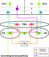
Schema of some neural circuitry of the thalamus, cortex and brainstem. Circuits showing first order (FO) and higher order (HO) connections of thalamocortical (TC) neurons (green cells) with neurons of the thalamic reticular nucleus (TRN; red cells) and cortical areas 1 (Ctx1) and 2 (Ctx2). Only the major thalamic-related input (layer 3/4) and output (layers 5 and 6) cortical laminae are shown. First order TC neurons receive driver afferents through ascending sensory pathways (green), whereas higher order TC neurons receive driver afferents through descending pathways (purple) from corticothalamic (CT) neurons in layer 5 (purple cell). Ascending and descending driver afferents have large axon terminals, whereas descending modulator afferents to TC and TRN neurons from CT neurons in layer 6 (blue cells) have small axon terminals. Note that TRN neurons can engage in closed-loop and/or open-loop circuits with TC neurons. Only brainstem modulator inputs (orange) from the pedunculopontine nucleus (PPN) are shown.
Figure 3
Figure 3
Schematic summary of different types of subnetworks in TRN. (A) TRN subnetworks identified according to their spatial distributions of electrically connected neurons through gap junctions (GJs) and connectivity with first order and/or higher order TC neurons. Elongated (circumscribed in orange) and broad (circumscribed in purple) GJ-related clusters of neurons (small gray ovals) are shown in TRN. (B) TRN subnetworks identified according to their state-dependent activity, location in TRN, and connectivity with first order thalamic nuclei. Neurons in dorsorostral TRN (orange) exhibit arousal-related activity whereas those in dorsocaudal TRN (blue) exhibit sleep-related activity. It is unclear whether this latter activity affects higher order sensory nuclei (LP and POl) in the thalamus. Abbreviations: AD, anterodorsal nucleus; dLGN, dorsal lateral geniculate nucleus; LP, lateral posterior nucleus; POl, posterior lateral nucleus. (C) TRN subnetworks identified according to their spatial distributions of neurons activated by glutamate corresponding to activation zones (AZs) and functional connectivity with first order and higher order TC neurons and driver-like-recipient thalamocortical/thalamostriatal (TC/TS) neurons. Elongated (circumscribed in orange) and broad (circumscribed in purple) AZ-related clusters of neurons (small gray ovals) are shown in TRN. (D) TRN subnetworks identified according to their mediation of interactions between groups of thalamic nuclei. Each group of interacting nuclei in the thalamus (shown on the right) is color coded. A possible distribution of neurons that mediate these interactions in TRN (shown on the left) is similarly color coded. Abbreviations: ILc, caudal intralaminar nuclei; ILr, rostral intralaminar nuclei; POm, posterior medial nucleus; TRN, thalamic reticular nucleus; VL, ventrolateral nucleus; VPM, ventroposterior medial nucleus.
Figure 4
Figure 4
Origins of driver-like afferents to some thalamic nuclei. Driver-like inputs originating from output nuclei of the basal ganglia (orange) are GABAergic whereas those originating from output layers of the superior colliculus (green) are glutamatergic. Abbreviations: dLGN, dorsal lateral geniculate nucleus; GPi, internal globus pallidus; IL, intralaminar nuclei; SNr, substantia nigra pars reticulata; VA, ventroanterior nucleus.
Figure 5
Figure 5
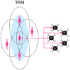
Individual neurons in the TRN are part of multiple overlapping neural clusters. Related to a particular type of thalamic projection neuron, individual reticular neurons in a neural cluster (e.g., central red cell of three cells shown underlying an activation zone (AZ; blue oval) shown on the left) are also part of other AZ-related neural clusters (outlined ovals) formed by neighboring reticular neurons, creating a dense subnetwork of multiple overlapping neural clusters in TRN. This anatomical arrangement would apply to both elongated and broad types of AZ-related clusters of reticular neurons (see text for details). Because each reticular neuron usually provides inhibitory inputs to several neighboring thalamocortical (TC) neurons (only five are shown on the right), the divergent outputs of reticular neurons suggest that single TC neurons can receive convergent inhibitory inputs from neurons in more than one neural cluster in TRN.

Similar articles

Cited by

References

    1. Acsády L. (2017). The thalamic paradox. Nat. Neurosci. 20, 901–902. 10.1038/nn.4583 - DOI - PubMed
    1. Ahrens S., Jaramillo S., Yu K., Ghosh S., Hwang G.-R., Paik R., et al. . (2015). ErbB4 regulation of a thalamic reticular nucleus circuit for sensory selection. Nat. Neurosci. 18, 104–111. 10.1038/nn.3897 - DOI - PMC - PubMed
    1. Alexander G. E., Crutcher M. D. (1990). Functional architecture of basal ganglia circuits: neural substrates of parallel processing. Trends Neurosci. 13, 266–271. 10.1016/0166-2236(90)90107-l - DOI - PubMed
    1. Alexander G. E., DeLong M. R., Strick P. L. (1986). Parallel organization of functionally segregated circuits linking basal ganglia and cortex. Annu. Rev. Neurosci. 9, 357–381. 10.1146/annurev.ne.09.030186.002041 - DOI - PubMed
    1. Aosaki T., Graybiel A. M., Kimura M. (1994a). Effect of the nigrostriatal dopamine system on acquired neural responses in the striatum of behaving monkeys. Science 265, 412–415. 10.1126/science.8023166 - DOI - PubMed

LinkOut - more resources