. 2018 Oct 22:10:332.
doi: 10.3389/fnagi.2018.00332. eCollection 2018.

Infection Augments Expression of Mechanosensing Piezo1 Channels in Amyloid Plaque-Reactive Astrocytes

Affiliations

Infection Augments Expression of Mechanosensing Piezo1 Channels in Amyloid Plaque-Reactive Astrocytes

María Velasco-Estevez et al. Front Aging Neurosci. .

Abstract

A defining pathophysiological hallmark of Alzheimer's disease (AD) is the amyloid plaque; an extracellular deposit of aggregated fibrillar Aβ1-42 peptides. Amyloid plaques are hard, brittle structures scattered throughout the hippocampus and cerebral cortex and are thought to cause hyperphosphorylation of tau, neurofibrillary tangles, and progressive neurodegeneration. Reactive astrocytes and microglia envelop the exterior of amyloid plaques and infiltrate their inner core. Glia are highly mechanosensitive cells and can almost certainly sense the mismatch between the normally soft mechanical environment of the brain and very stiff amyloid plaques via mechanosensing ion channels. Piezo1, a non-selective cation channel, can translate extracellular mechanical forces to intracellular molecular signaling cascades through a process known as mechanotransduction. Here, we utilized an aging transgenic rat model of AD (TgF344-AD) to study expression of mechanosensing Piezo1 ion channels in amyloid plaque-reactive astrocytes. We found that Piezo1 is upregulated with age in the hippocampus and cortex of 18-month old wild-type rats. However, more striking increases in Piezo1 were measured in the hippocampus of TgF344-AD rats compared to age-matched wild-type controls. Interestingly, repeated urinary tract infections with Escherichia coli bacteria, a common comorbidity in elderly people with dementia, caused further elevations in Piezo1 channel expression in the hippocampus and cortex of TgF344-AD rats. Taken together, we report that aging and peripheral infection augment amyloid plaque-induced upregulation of mechanoresponsive ion channels, such as Piezo1, in astrocytes. Further research is required to investigate the role of astrocytic Piezo1 in the Alzheimer's brain, whether modulating channel opening will protect or exacerbate the disease state, and most importantly, if Piezo1 could prove to be a novel drug target for age-related dementia.

Keywords: Alzheimer’s disease; Piezo1; TgF344-AD rats; amyloid plaques; astrocytes; dentate gyrus; mechanosensitive ion channel; urinary tract infection.


Figures

FIGURE 1
FIGURE 1
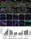
Piezo1 channels are highly expressed by neurons in the optic tract, corpus callosum, and arbor vitae of the cerebellum. Coronal sections of the 5-week old rat brain were cut at –3.3 mm (A) and –10.8 mm (B) with respect to Bregma and immunofluorescently stained for Piezo1 (N-terminal antibody) (n = 4), scale bar = 4,000 μm. The red arrow points to the optic tract in the right hemisphere and the white arrow points to the arbor vitae of the Ansiform lobule Crus 1 of the cerebellum. 5-week old rat brains were also dissected into four parts for Western blotting (WB), i.e., cerebellum (Crb), hippocampus (Hip), cerebral cortex (Ctx) and brainstem/thalamus (BS/T). WB were run for Piezo1 using an N-terminal binding antibody (C,D) from Santa Cruz (n = 3) and a C-terminal binding antibody (E,F) from Abcam (n = 3). The hippocampus consistently showed the lowest levels of Piezo1 protein expression.
FIGURE 2
FIGURE 2
Piezo1 channel expression in the adult rat brain. (A) The brains of adult (12 month) Fisher rats were sectioned in the sagittal plane (lateral –1.9 mm) and stained for (B) Piezo1 (N-terminal antibody), (C) DAPI, and (D) GFAP (n = 5). Images were captured using a 20× magnification objective on an Axio Scan.Z1 slide scanner (Zeiss, Germany) and montaged in ZEN software. 20× magnification images of the hippocampal (E) CA1 (scale bar = 150 μm), (F) CA3 and (G) dentate gyrus (DG) show that Piezo1 expression is greater in pyramidal and granule neurons compared to GFAP-positive astrocytes. In the cerebral cortex, (H) layer V pyramidal neurons (LV Ctx) appear to express more Piezo1 channels than (I) prefrontal cortical (PFC) neurons. In the cerebellum (J), Purkinje neuron cell bodies and axons (arbor vitae) express higher levels of Piezo1 compared to neurons in the granule and molecular cell layers. (K) The dorsal midbrain (DM) expresses moderate levels of Piezo1, whereas (L) epithelial cells of the choroid plexus (CP) express greater amounts of Piezo1 protein. It was also noted that white matter tracts in (M) the thalamus (Th) and (N) striatum (Str) express more Piezo1 than gray matter areas of these brain regions. The areas depicted in images (E–N) are shown as yellow boxes in (C). Relative fluorescence intensity (arbitrary fluorescence units, a.f.u.) of Piezo1 staining in each brain region was quantified (O). Axonal tracts including the cerebellar arbor vitae, hippocampal fimbriae, cingulum and corpus callosum, as well as the medulla and pons of the brainstem, had the highest expression of Piezo1 protein.
FIGURE 3
FIGURE 3
1-42 CMM upregulates Piezo1 expression in cortical astrocyte cultures. To check cellular localisation of Piezo1 in the brain, enriched astrocyte cultures (A–D) and cortical neuronal cultures (E–H) from newborn mice were stained for (A,E) Piezo1 (N-terminal antibody) and (C) GFAP or (G) neurofilament H (NFH), respectively (n = 3). Piezo1 co-localized with NFH staining in neurons (H) but was almost completely absent from astrocytes under basal conditions (D). To investigate if neuroinflammatory stimuli can induce Piezo1 expression in astrocytes, mouse microglia were exposed to human Aβ1-42 (Invitrogen, United Kingdom) for 24 h and the inflammatory media was collected. Enriched mouse astrocyte cultures were then exposed to this Aβ1-42 CMM for 48 h. Astrocytes were immunofluorescently stained for (I) Piezo1 (C-terminus antibody), (J) DAPI, (K) GFAP, and (L) is the merge of each channel. Scale bar = 20 μm. Exposure to Aβ1-42 CMM upregulated Piezo1 protein expression in approximately 30–40% of astrocytes (M,N). Astrocytes were also treated for 24 h with either 5 μM hydrogen peroxide (H2O2) or a cytokine cocktail consisting of TNF-α (10 ng/mL) and IL17A (250 ng/mL). Neither H2O2 nor TNF-α/IL17A increased Piezo1 expression indicating that Piezo1 upregulation is not a common response to all cell stressors but specific to certain inflammatory stimuli including Aβ1-42 and factors released by reactive amyloid-stimulated microglia. Data are represented as mean ± SEM (n = 8). A repeated measures one-way ANOVA with Holm-Sidak post hoc test was performed. Represents a statistically significant difference (p < 0.05) from control (Ctrl), TNF-α/IL17A, and H2O2 treatment groups.
FIGURE 4
FIGURE 4
1-42 pathology in vivo upregulates Piezo1 expression in astrocytes surrounding amyloid plaques. 18-Month old TgF344-AD rat brains were immunofluorescently stained for (A) conformation-specific Aβ1-42, (B) GFAP, (C) Piezo1 (N-terminal antibody), and (D) DAPI. Scale bar = 40 μm. (E) High magnification (100× objective) z-stack projections of prefrontal cortex amyloid plaques reveal an upregulation of GFAP in reactive astrocytes surrounding the amyloid plaque. Piezo1 protein is also upregulated in a proportion of astrocytes in and around the core of the plaque. Piezo1 appears localized predominantly to the perinuclear compartment of Aβ1-42 reactive astrocytes. (F) Astrocytic Piezo1 fluorescence intensity was calculated for astrocytes inside the amyloid plaque versus astrocytes located 200–500 μm away from any noticeable plaque deposits. Piezo1 fluorescence intensity was averaged for six astrocytes “inside” and six astrocytes “outside” the plaque and 25 images from the frontal cortex of 18 m TG rats (n = 6) were analyzed. There was no correlation (Pearson r = 0.468) between Piezo1 expression inside versus outside amyloid plaques. (G) However, on average there was a 190% increase in Piezo1 expression in astrocytes within the plaque core (Inside) versus astrocytes at least 200 μm away from the edge of any amyloid plaque (Outside). A two-tailed paired t-test was performed. Represents a statistically significant difference (p < 0.001) inside versus outside of the plaque.
FIGURE 5
FIGURE 5
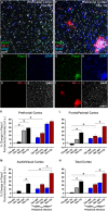
Mechanosensing Piezo1 channel expression increases in the cerebral cortex of TgF344-AD rats with a urinary tract infection (UTI). Sagittal sections (lateral –1.9 mm) of wild-type (WT) Fisher and TgF344-AD rat brains were immunofluorescently stained for Piezo1 (N-terminal antibody), conformation-specific Aβ1-42, GFAP, and DAPI. Shown are representative images of the prefrontal cortex of an 18-month WT rat (A–E) and an 18-month TgF344-AD (18 m TG) (F–J). Scale bar = 150 μm. Elevations in Piezo1 channel expression with age, Alzheimer’s disease (AD) pathology and peripheral infection were measured in three distinct areas of the cerebral cortex, i.e., the prefrontal cortex (K), the frontoparietal cortex (L), and the audiovisual cortex (M). In addition, Piezo1 expression was averaged over all three regions to illustrate changes in channel expression in the total cerebral cortex (N). Data were normalized to 12 month wild-type (12m WT) values for each cortical region and are represented as the mean ± SEM of the percentage change from the 12m WT groups. Three-way ANOVAs with Holm-Sidak post hoc tests were performed to test for interactions between age, genotype, and peripheral infection. Represents a change > 20% and a p-value < 0.01. There were 5–6 rats per group and 2–3 brain sections per animal. Ten regions of interest (ROI) were analyzed per cortical region per section, i.e., 100–180 ROIs analyzed per group.
FIGURE 6
FIGURE 6
Aging, Aβ1-42 pathology and peripheral infection upregulate Piezo1 expression in the hippocampus of TgF344-AD rats. Sagittal sections (lateral -1.9 mm) of WT Fisher and TgF344-AD rat brains were immunofluorescently stained for Piezo1 (N-terminal antibody), conformation-specific Aβ1-42, GFAP, and DAPI. Shown are representative images of the hippocampus of an 18-month wild-type (18m WT) rat (A–E), an 18-month TgF344-AD 18m TG (F–J), an 18-month WT with infection (18m iWT) (K–O), and an 18-month TgF344-AD with infection (18m iTG) (P–T). TgF344-AD rats with and without an infection show clear upregulations of GFAP (I,S) in hippocampal areas with dense amyloid deposition (H,R), particularly the hilus, the outer molecular layer of the dentate gyrus, and the stratum oriens of the CA1. Moreover, amyloid plaques surrounded by a dense meshwork of GFAP-positive astrocytes generally showed concomitant upregulations of Piezo1 expression, particularly in 18m TG rats with or without an infection (G,Q). Next, changes in Piezo1 expression with age, infection and genotype were quantified in the neuronal cell body layers of the dentate gyrus (U), CA3 (V), and CA1 (W). Piezo1 expression increased from 12- to 18-months in WT rats in the DG and CA1, but not CA3 neurons. A much more potent trigger of Piezo1 expression in the hippocampus, however, was genotype with TgF344-AD rats expressing much larger levels of Piezo1 in the DG (U), CA3 (V), and CA1 (W) at 12-months of age. There were no further increases in Piezo1 expression in 18-month old TgF344-AD rats. However, peripheral infection caused a significant increase in Piezo1 expression in the dentate gyrus (U) of TgF344-AD rats (18m iTG). Data were normalized to 12m WT values for each hippocampal region and are represented as the mean ± SEM of the percentage change from the 12m WT groups. Three-way ANOVAs with Holm-Sidak post hoc tests were performed to test for interactions between age, genotype, and peripheral infection. Represents a change of > 20% and a p-value < 0.01. There were 5–6 rats per group and 2–3 brain sections per animal. Ten ROI were analyzed per hippocampal region per section, i.e., 100–180 ROIs analyzed per group.
FIGURE 7
FIGURE 7
Piezo1 expression in the hippocampal dentate gyrus correlates with GFAP in aged rats with peripheral infection and strongly correlates with amyloid plaques in TgF344-AD rats. Sagittal brain sections were triple-stained for Piezo1, GFAP and conformation-specific Aβ1-42 and Pearson correlations (r values) were performed to measure changes in Piezo1 channel expression in astrocytes (GFAP vs. Piezo1) with age and peripheral infection in WT (A–D) and TgF344-AD (E–H) rats. There was a moderate Pearson correlation between GFAP and Piezo1 in the infected 18-month WT (r = 0.523) and infected 18-month TgF344-AD rats (r = 0.519). However, the linear regression (R2) values were relatively weak for both groups suggesting that the GFAP fluorescence intensity is not a good predictor of Piezo1 channel expression. Next, Pearson correlations were performed to measure changes in GFAP expression around amyloid plaques (GFAP vs. Aβ1-42) in the dentate gyrus with age and peripheral infection in TgF344-AD rats (I–L). There was a moderate Pearson correlation between GFAP and Aβ1-42 in the infected 18-month old TgF344-AD rats (r = 0.586) but a relatively weak linear regression (R2) value suggesting that Aβ1-42 fluorescence intensity is not a good predictor of GFAP expression. Finally, Pearson correlations were performed to measure changes in Piezo1 channel expression in and around amyloid plaques (Aβ1-42 vs. Piezo1) in the dentate gyrus with age and peripheral infection in TgF344-AD rats (M–P). There were strong Pearson correlations between Aβ1-42 and Piezo1 in 12-month old TgF344-AD rats (r = 0.917), 12-month old TgF344-AD rats with peripheral infection (r = 0.736), 18-month old TgF344-AD rats (r = 0.770), and 18-month old TgF344-AD rats with peripheral infection (r = 0.856). In addition, the high linear regression (R2) values for each group (M–P) suggests that Aβ1-42 fluorescence intensity is a good predictor of Piezo1 channel expression in the dentate gyrus of TgF344-AD rats.

Similar articles

Cited by

References

    1. Arnsten A. F., Li B. M. (2005). Neurobiology of executive functions: catecholamine influences on prefrontal cortical functions. Biol. Psychiatry 57 1377–1384. 10.1016/j.biopsych.2004.08.019 - DOI - PubMed
    1. Bae C., Sachs F., Gottlieb P. A. (2015). Protonation of the human PIEZO1 ion channel stabilizes inactivation. J. Biol. Chem. 290 5167–5173. 10.1074/jbc.M114.604033 - DOI - PMC - PubMed
    1. Barbas H., Zikopoulos B. (2007). The prefrontal cortex and flexible behavior. Neuroscientist 13 532–545. 10.1177/1073858407301369 - DOI - PMC - PubMed
    1. Bavi N., Nikolaev Y. A., Bavi O., Ridone P., Martinac A. D., Nakayama Y., et al. (2017). “Principles of mechanosensing at membrane interface,” in The Biophysics of Cell Membranes, 19 Edn, eds Epand R. M., Ruysschaert J.-M. (Berlin: Springer Series in Biophysics Springer; ), 85–119. 10.1007/978-981-10-6244-5_4 - DOI
    1. Blumenthal N. R., Hermanson O., Heimrich B., Shastri V. P. (2014). Stochastic nanoroughness modulates neuron-astrocyte interactions and function via mechanosensing cation channels. Proc. Natl. Acad. Sci. U.S.A. 111 16124–16129. 10.1073/pnas.1412740111 - DOI - PMC - PubMed

LinkOut - more resources