. 2018 Oct 23:9:2301.
doi: 10.3389/fimmu.2018.02301. eCollection 2018.

A Metalloprotease Homolog Venom Protein From a Parasitoid Wasp Suppresses the Toll Pathway in Host Hemocytes

Affiliations

A Metalloprotease Homolog Venom Protein From a Parasitoid Wasp Suppresses the Toll Pathway in Host Hemocytes

Zhe Lin et al. Front Immunol. .

Abstract

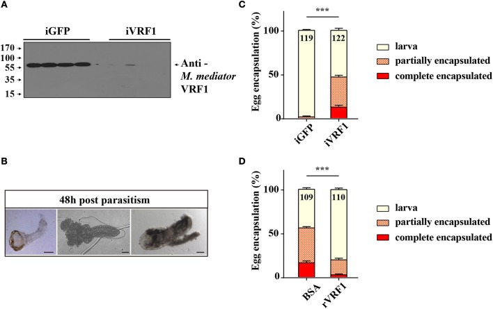
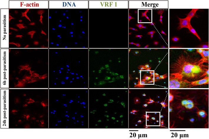
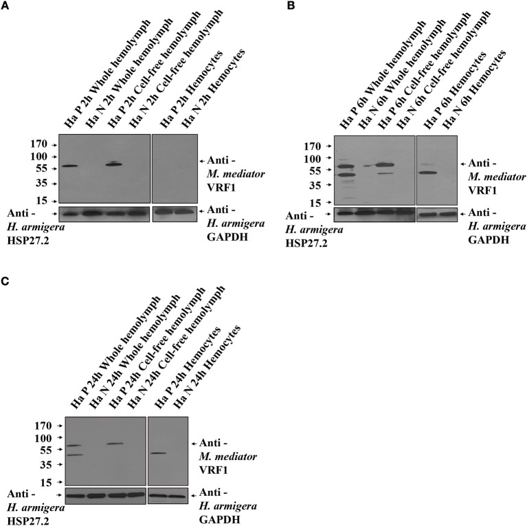
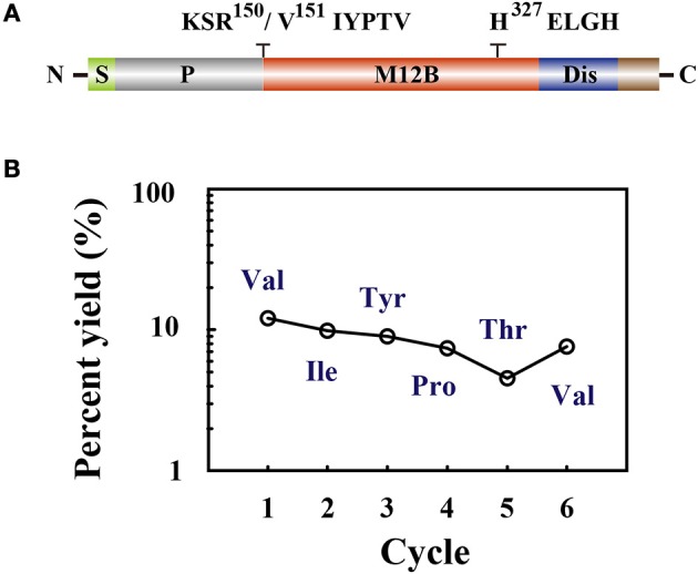
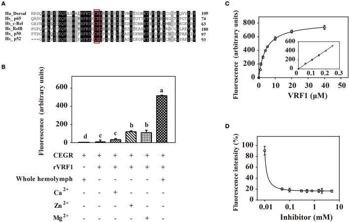
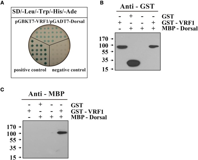
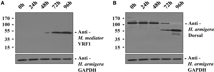
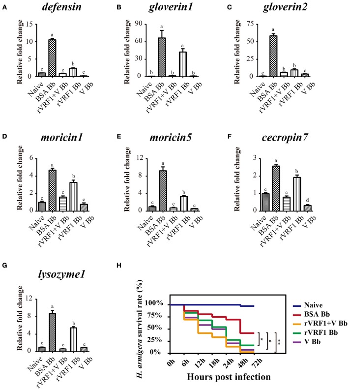
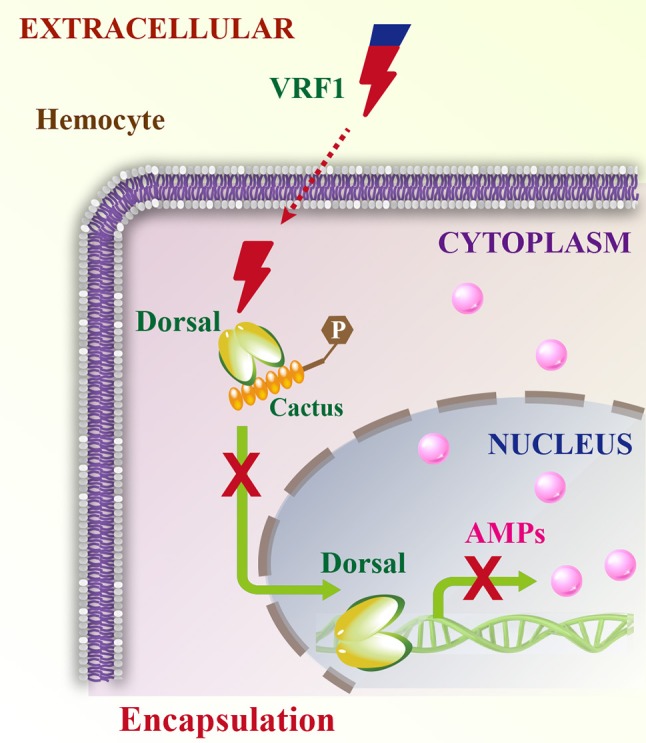
Parasitoid wasps depend on a variety of maternal virulence factors to ensure successful parasitism. Encapsulation response carried out by host hemocytes is one of the major host immune responses toward limiting endoparasitoid wasp offspring production. We found that VRF1, a metalloprotease homolog venom protein identified from the endoparasitoid wasp, Microplitis mediator, could modulate egg encapsulation in its host, the cotton bollworm, Helicoverpa armigera. Here, we show that the VRF1 proenzyme is cleaved after parasitism, and that the C-terminal fragment containing the catalytic domain enters host hemocytes 6 h post-parasitism. Furthermore, using yeast two-hybrid and pull-down assays, VRF1 is shown to interact with the H. armigera NF-κB factor, Dorsal. We also show that overexpressed of VRF1 in an H. armigera cell line cleaved Dorsal in vivo. Taken together, our results have revealed a novel mechanism by which a component of endoparasitoid wasp venom interferes with the Toll signaling pathway in the host hemocytes.

Keywords: Helicoverpa armigera; Microplitis mediator; metalloprotease; toll pathway; venom.


Figures

Figure 1
Figure 1
The interaction between M. mediator VRF1 and egg encapsulation. (A) The efficiency of M. mediator VRF1 RNA interference (iVRF1) was confirmed by immunoblot analysis using VRF1 antibody (1:5,000). Three days after injecting dsRNA, venom reservoir was excised from individual females. Venom solution from each sample was diluted to a final concentration of 0.2 venom reservoir equivalents. iGFP injected wasps were used as the control. (B) Wasp eggs or larvae were dissected 48 h post-parasitism by visualizing via light microscopy. Wasp offspring were divided into three types based on their phenotype: larva, partially encapsulated, or completely encapsulated (images from left to right). Scale bar, 20 μm. (C) Egg encapsulation rate (%) measured after depletion of VRF1 (iVRF1) when compared with dsGFP-injected wasps (iGFP). Each group contained 10 RNAi wasps that individually parasitized 30 s instar H. armigera larvae. Each RNAi treatment contained five groups. Data represent the numbers of recovered offspring analyzed. Statistical significance of the egg encapsulation rate was analyzed using the Chi-square test. Error bars represent the standard error of the mean (SEM). Statistically significant differences between groups were evaluated by t-test (Mann-Whitney nonparametric test), ***p < 0.001. (D) Egg encapsulation rate (%) was partially rescued following rVRF1 injection. rVRF1, second instar larvae of H. armigera were injected rVRF1 (2 × 10⁻⁷ μg rVRF1/larva), 24 h later, were parasitized by VRF1 depleted wasps. BSA, second instar larvae of H. armigera were injected the same amount of 200 nL BSA, 24 h latter, were parasitized by VRF1 depleted wasps. Each group consisted of 10 RNAi injected wasps that individually parasitized 30 hosts (one wasp individually parasitized three hosts). Each treatment contained five groups. Data represent the numbers of recovered offspring analyzed Statistical significance of the egg encapsulation rate was analyzed using the Chi-square test. Error bars represent the standard error of the mean (SEM). Statistically significant differences between groups were evaluated by t-test (Mann-Whitney nonparametric test), ***p < 0.001.
Figure 2
Figure 2
VRF1 localized near host hemocyte nucleus after parasitism. Confocal micrographs of hemocytes from second instar H. armigera larvae, no parasitism, 6 h post-parasitism, and 24 h post-parasitism. F-actin and hemocytes DNA were visualized with Phalloidin-Alexa Fluor 594 (red) and Hoechst 33342 (blue) using Image-IT cell labeling kit. VRF1 was detected using a specific rabbit polyclonal antibody (green). The white boxed areas are enlarged in the right panel and show the hemocytes cluster. Scale bar, 20 μm.
Figure 3
Figure 3
VRF1 was cleaved into two parts after entering natural host's hemocoel. (A) Immunoblot analysis of whole hemolymph, cell-free hemolymph, and hemocytes from second instar H. armigera larvae at 2 h post-parasitism (P 2h) and unparasitized second instar H. armigera larvae (N 2h), using VRF1 antibody. (B) Immunoblot analysis of whole hemolymph, cell-free hemolymph and hemocytes from second instar H. armigera larvae 6 h post-parasitism (P 6h) and unparasitized second instar H. armigera larvae (N 6h), using VRF1 antibody. (C) Immunoblot analysis of whole hemolymph, cell-free hemolymph, and hemocytes from second instar H. armigera larvae at 24 h post-parasitism (P 24h) and unparasitized second instar H. armigera larvae (N 24h), using VRF1 antibody. HSP27.2 (H. armigera heat shock protein 27.2 kDa) antibody (1:5,000) was used as the loading control for the whole and cell free hemolymph samples. GAPDH (H. armigera glyceraldehyde-3-phosphate dehydrogenase) antibody (1:5,000) was used as the loading control for hemocytes.
Figure 4
Figure 4
(A) The domain structure is indicated as signal peptide (S), prodomain (P), metalloprotease 12B domain (M12B), and disintegrin-like domain (Dis). Amino acid sequences KSR150/V151IYPTV showed that cleavage occurred after R150. The conserved zinc-metalloprotease signature sequences H327ELGH shown in their relative positions. (B) Amino acid sequences of the collected peptides (45 kDa) were determined by Edman degradation, which was carried out for 6 cycles. The peptide sequence revealed was Val-Ile-Tyr-Pro-Thr-Val.
Figure 5
Figure 5
rVRF1 could hydrolyze artificial substrate containing the putative cleavage site. (A) Sequence alignment of H. armigera Dorsal and human Rel family proteins. Identical or similar residues are highlighted in black and gray colors respectively. Boxed sequences indicate the conserved Cys-Glu residues. (B) Fluorescence assay of rVRF1 in different reactions. To test the enzymatic activity of rVRF1, the fluorogenic substrate peptides comprising the sequence DABCYL-Cys-Glu-Gly-Arg-Ser-Ala-EDANS-NH2 (CEGR) were synthesized. Substrate CEGR at 0.05 μg/μL was incubated with rVRF1 at 5 μM at room temperature for 30 min. The final reaction volume of 200 μL contained 50 mM Tri-HCl, 150 mM NaCl, 0.2 mM NaN3 with 5 μL whole hemolymph from second instar H. armigera larvae or 5 mM CaCl2, ZnCl2, and MgCl2 respectively. Fluorescence was measured using a microplate reader (SpectraMax i3, Molecular Devices) with endpoint assay set at excitation wavelengths of 335 nm and emission wavelengths of 493 nm. Error bars represent the means ± SEM from three replicates. Different letters above a given bar represents significant difference in Tukey's multiple comparison test (p < 0.05). (C) rVRF1 enzymatic activity assay. Reactions contained 0.05 μg/μL substrate CEGR (DABCYL-Cys-Glu-Gly-Arg-Ser-Ala-EDANS-NH2), the indicated amount of rVRF1 (0, 1, 2, 3, 4, 5, 10, 20, and 40 μM) and 5 μL whole hemolymph from second instar H. armigera larvae. After incubation for 30 mins at room temperature, fluorescence was measured using a microplate reader (SpectraMax i3, Molecular Devices) with endpoint assay set for excitation at 335 nm and emission detection at 493 nm. The inset shows an enlargement of the plot depicting the values for low concentrations of rVRF1. Data were fitted to the Michaelis-Menten equation using GraphPad Prism (version 6.0). (D) Assay of rVRF1 inhibition curve by 1,10-phenanthroline. Substrate CEGR (DABCYL-Cys-Glu-Gly-Arg-Ser-Ala-EDANS-NH2) at 0.05 μg/μL, 5 μM rVRF1, 5 μL whole hemolymph of second instar H. armigera larvae and increasing amounts of 1,10-phenanthroline (Sigma) at 0.01, 0.05, 0.1, 0.3, 0.5, 0.8, 1.5, 2, 5 mM were mixed and incubated at room temperature for 30 mins. Fluorescence was measured as described in Figure 5C.
Figure 6
Figure 6
M. mediator VRF1 binds H. armigera Dorsal. (A) Yeast two hybrid assay between M. mediator VRF1 and H. armigera Dorsal in high-stringency binding test media of SD-Leu-Trp-His-Ade with X-α-gal. pGBKT7-53 and pGADT7-T were cotransformed as positive controls, while pGBKT7-Lam and pGADT7-T were cotransformed as negative controls. (B) In vitro GST-pull-down assay of recombinant MBP-Dorsal and GST-VRF1. Immunoblot analysis of eluted proteins from Glutathione Sepharose 4B using GST antibody. (C) Immunoblot analysis of eluted proteins from Glutathione Sepharose 4B using MBP antibody.
Figure 7
Figure 7
Overexpression of M. mediator VRF1 in an H. armigera cell line. (A) Immunoblot analysis of IOZCAS-Ha-I cells after transfection with pIZT/VRF1151−483 post 0, 24, 48, 72, and 96 h using M. mediator VRF1 antibody. GAPDH (H. armigera glyceraldehyde-3-phosphate dehydrogenase) antibody (1:5,000) was used as the loading control for cells. (B) Immunoblot analysis of IOZCAS-Ha-I cells after transfection with pIZT/VRF1151−483 post 0, 24, 48, 72, and 96 h using H. armigera Dorsal antibody. GAPDH (H. armigera glyceraldehyde-3-phosphate dehydrogenase) antibody (1:5,000) was used as the loading control for cells.
Figure 8
Figure 8
The expression of host AMP genes and survival rate analysis by injection of rVRF1 and venom. (A–G) The mRNA expression level of several AMP genes were measured with hemocytes collected from second instar H. armigera larvae in the different treatment groups. (A) defensin; (B) gloverin1; (C) gloverin2; (D) moricin1; (E) moricin5; (F) cecropin7; (G) lysozyme1. Naïve, untreated second instar larvae of H. armigera. BSA Bb, second instar H. armigera larvae injected 200 nL of BSA (2× 10⁻⁷ μg BSA/larva), and one day latter infected with B. bassiana (2 × 10³ conidia/larva). rVRF1 Bb, larvae were injected 200 nL of rVRF1 (2× 10⁻⁷ μg rVRF1/larva), and 1 day latter, was infected with B. bassiana (2 × 10³ conidia/larva). V Bb, larvae were injected 200 nL of venom (0.05 w.e. venom/larva), and 1 day latter infected with B. bassiana (2 × 10³ conidia/larva). rVRF1+V Bb, larvae were injected 200 nL of 2 × 10⁻⁷ μg rVRF1 and 0.05 w.e. venom/larva, and 1 day latter challenged with B. bassiana (2 × 10³ conidia/larva). Error bars represent the means ± SEM from four replicates. Differences between treatments were compared by one-way ANOVA followed by Tukey's test for multiple comparisons. Different letters above a given bar represent significant difference among the different injection groups (p < 0.05). (H) Survival rate analysis of H. armigera after injection and infection experiments. Each group contained 72 s instar larvae and the experiment had three replications. Data were analyzed using the Kaplan-Meier method (*p < 0.05, **p < 0.01).
Figure 9
Figure 9
Schematic model of VRF1 action. Venom protein VRF1 (cleaved) from M. mediator interacts with Dorsal to suppress the transcription of host cell AMP genes.

Similar articles

Cited by

References

    1. Asgari S, Rivers DB. Venom proteins from endoparasitoid wasps and their role in host-parasite interactions. Annu Rev Entomol. (2011) 56:313–35. 10.1146/annurev-ento-120709-144849 - DOI - PubMed
    1. Strand MR, Pech LL. Immunological basis for compatibility in parasitoid-host relationships. Annu Rev Entomol. (1995) 40:31–56. 10.1146/annurev.en.40.010195.000335 - DOI - PubMed
    1. Harvey JA, Poelman EH, Tanaka T. Intrinsic inter- and intraspecific competition in parasitoid wasps. Annu Rev Entomol. (2013) 58:333–51. 10.1146/annurev-ento-120811-153622 - DOI - PubMed
    1. Russo J, Dupas S, Frey F, Carton Y, Brehelin M. Insect immunity: early events in the encapsulation process of parasitoid (Leptopilina boulardi) eggs in resistant and susceptible strains of Drosophila. Parasitology (1996) 112:135–42. 10.1017/S0031182000065173 - DOI - PubMed
    1. Mortimer NT, Kacsoh BZ, Keebaugh ES, Schlenke TA. Mgat1-dependent N-glycosylation of membrane components primes Drosophila melanogaster blood cells for the cellular encapsulation response. PLoS Pathog. (2012) 8:e1002819. 10.1371/journal.ppat.1002819 - DOI - PMC - PubMed

Publication types

LinkOut - more resources