. 2018 Oct 22:9:2408.
doi: 10.3389/fimmu.2018.02408. eCollection 2018.

Incubation of Immune Cell Grafts With MAX.16H5 IgG1 Anti-Human CD4 Antibody Prolonged Survival After Hematopoietic Stem Cell Transplantation in a Mouse Model for Fms Like Tyrosine Kinase 3 Positive Acute Myeloid Leukemia

Affiliations

Incubation of Immune Cell Grafts With MAX.16H5 IgG1 Anti-Human CD4 Antibody Prolonged Survival After Hematopoietic Stem Cell Transplantation in a Mouse Model for Fms Like Tyrosine Kinase 3 Positive Acute Myeloid Leukemia

Nadja Hilger et al. Front Immunol. .

Abstract

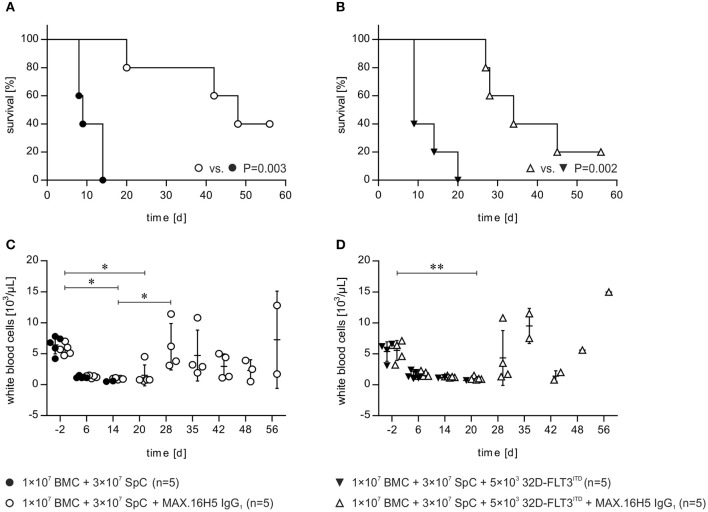
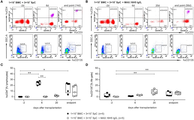
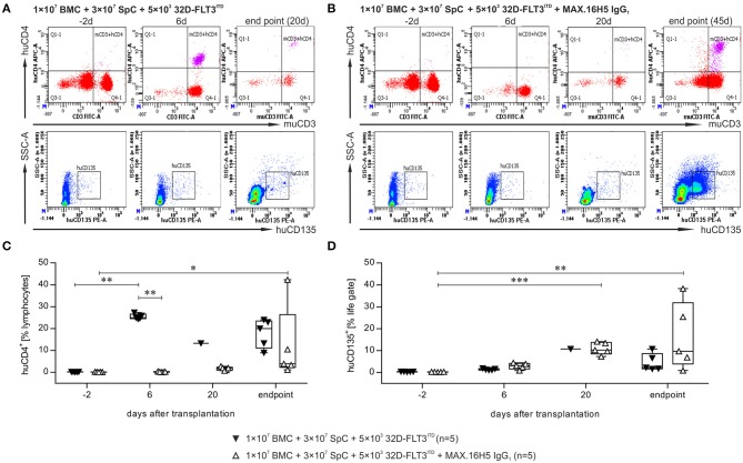
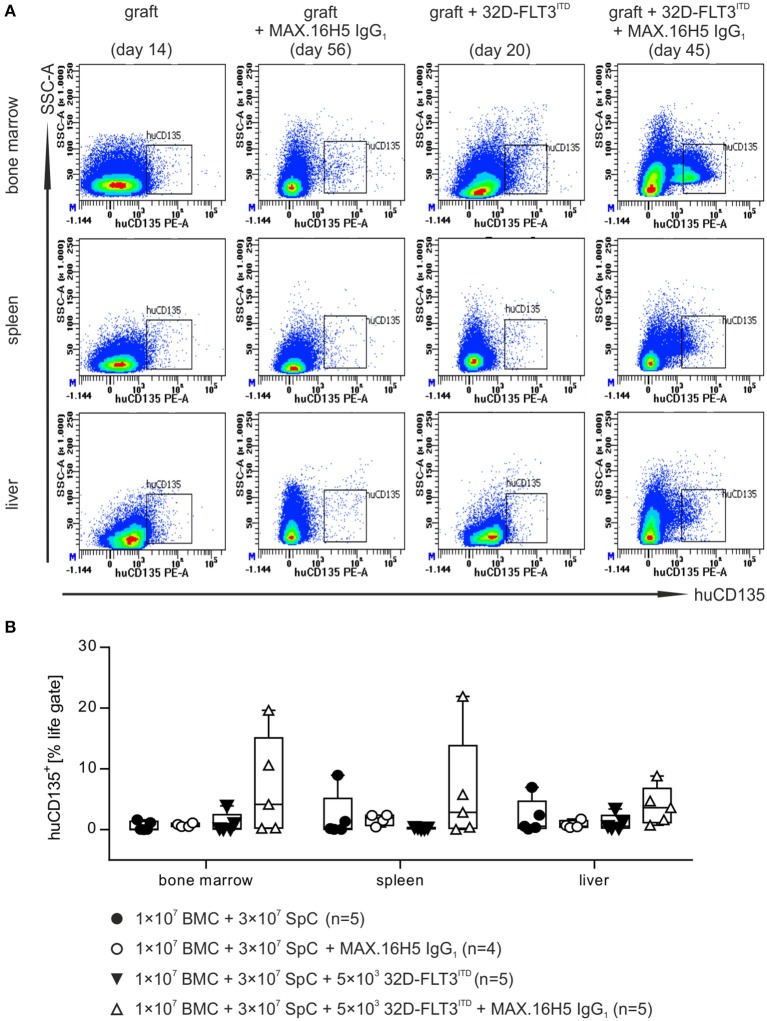
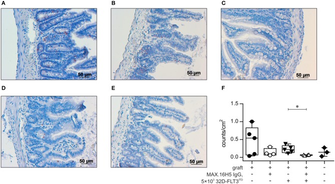
Despite the constant development of innovative therapeutic options for hematological malignancies, the gold-standard therapy regimen for curative treatment often includes allogeneic hematopoietic stem cell transplantation (HSCT). The graft-vs.-leukemia effect (GVL) is one of the main therapeutic goals that arises from HSCT. On the other hand, graft-vs.-host disease (GVHD) is still one of the main and most serious complications following allogeneic HSCT. In acute myeloid leukemia (AML), HSCT together with high-dose chemotherapy is used as a treatment option. An aggressive progression of the disease, a decreased response to treatment, and a poor prognosis are connected to internal tandem duplication (ITD) mutations in the Fms like tyrosine kinase 3 (FLT3) gene, which affects around 30% of AML patients. In this study, C3H/HeN mice received an allogeneic graft together with 32D-FLT3ITD AML cells to induce acute GVHD and GVL. It was examined if pre-incubation of the graft with the anti-human cluster of differentiation (CD) 4 antibody MAX.16H5 IgG1 prevented the development of GVHD and whether the graft function was impaired. Animals receiving grafts pre-incubated with the antibody together with FLT3ITD AML cells survived significantly longer than mice receiving untreated grafts. The observed prolonged survival due to MAX.16H5 incubation of immune cell grafts prior to transplantation may allow an extended application of additional targeted strategies in the treatment of AML.

Keywords: 32D-FLT3ITD; C3H/HeN; acute myeloid leukemia; anti-human CD4 antibody MAX.16H5; graft-vs.-host disease; graft-vs.-leukemia; hematopoietic stem cell transplantation.


Figures

Figure 1
Figure 1
Short-term pre-incubation of bone marrow and spleen cell grafts with MAX.16H5 IgG1 antibody prolonged survival of C3H/HeN recipient mice after allogeneic transplantation either with or without co-transplantation of 32D-FLT3ITD AML cells. All C3H/HeN mice received 1 × 10⁷ bone marrow cells (BMC) and 3 × 10⁷ spleen cells (SpC) as an allogeneic transplant in these experiments. (A) Survival of recipient mice after allogeneic transplantation. The grafts were pre-incubated either with MAX.16H5 IgG1 or without antibody before administration to the recipients. C3H/HeN animals receiving a MAX.16H5 IgG1 pre-incubated graft (n = 5, open circles) showed a significantly prolonged survival compared to animals receiving an untreated graft (n = 5, closed circles, P = 0.003; Log-Rank test). (B) Survival of C3H/HeN recipient mice receiving short-term pre-incubated (with or without MAX.16H5 IgG1 antibody) grafts together with 5 × 10³ 32D-FLT3ITD AML cells. C3H/HeN animals receiving a MAX.16H5 IgG1 pre-incubated graft together with 32D-FLT3ITD cells (n = 5, open triangle) showed a statistically significant prolonged survival compared to animals receiving an antibody untreated graft (n = 5, closed triangle, P = 0.002; Log-Rank test). (C) Reconstitution of white blood cells in the blood of C3H/HeN mice which received either MAX.16H5 IgG1 pre-incubated grafts (n = 5, open circle) or grafts without prior antibody incubation (n = 5, closed circles). (D) White blood cell counts in the blood of C3H/HeN mice receiving bone marrow and spleen cell grafts either pre-incubated with MAX.16H5 IgG1 antibody (n = 5, open triangle) or without antibody pre-incubation (n = 5, closed triangles) is depicted. The blood sample data in (C,D) were obtained by electrical impedance measurement using a hematology analyzer. The error bars represent time-point specific means ± standard deviations. Nemenyi post-hoc tests with Bonferroni correction were used for comparative statistics. *P < 0.05, **P < 0.01.
Figure 2
Figure 2
Flow cytometric analysis of the immune reconstitution (murine CD3+/human CD4+) as well as tumor growth (human CD135+) after transplantation of bone marrow and spleen cell grafts either pre-incubated with MAX.16H5 IgG1 or without antibody pre-treatment. All recipient animals received 1 × 10⁷ bone marrow cells (BMC) and 3 × 10⁷ splenocytes (SpC) in these experiments. (A,B) Representative plots obtained from flow cytometric analyses after transplantation of (A) one animal with untreated or (B) one animal with MAX.16H5 IgG1 pre-treated grafts at different time points (in (A): day −2, day 6, endpoint; in (B): day −2, day 6, day 20, endpoint) using specific antibodies against murine CD3/human CD4 or human CD135. In the lower rows, human CD135+ signals are depicted together with side-scattered light signals. (C) Human CD4+ events as percentages of lymphocyte gates in the blood samples of recipient mice. Every symbol indicates one animal at a defined time point. Engraftment and proliferation of human CD4+ cells were detected in five out of five animals receiving an untreated graft and in three out of five animals receiving the MAX.16H5 IgG1 pre-incubated grafts. The immune cell engraftment was delayed in animals receiving the MAX.16H5 IgG1 pre-incubated grafts. (D) Human CD135+ control staining of blood samples on labeled time points. (C,D) Nemenyi tests with Bonferroni correction computed the comparative statistics. *P < 0.05, **P < 0.01.
Figure 3
Figure 3
Flow cytometric analysis of the immune reconstitution (murine CD3+/human CD4+) as well as tumor growth (human CD135+) after transplantation of bone marrow and spleen cell grafts pre-incubated with MAX.16H5 IgG1 or without antibody pre-treatment in co-transplantation experiments with 5 × 10³ 32D-FLT3ITD cells. All recipient animals received 1 × 10⁷ bone marrow cells (BMC) and 3 × 10⁷ splenocytes (SpC) in these experiments. (A,B) Representative plots of flow cytometric analyses after transplantation of (A) one animal with untreated or (B) one animal with MAX.16H5 IgG1 pre-treated grafts at different time points [in (A): day −2, day 6, endpoint; in (B): day −2, day 6, day 20, endpoint] using specific antibodies for murine CD3+/human CD4+ or human CD135+. In the lower rows, human CD135+ signals are depicted together with side-scattered light signals. (C) Human CD4+ events as percentages of lymphocyte gates in the blood samples of recipient mice. Every symbol indicates one animal at a defined time point. Engraftment and proliferation of human CD4+ cells were detected in five out of five animals receiving an antibody untreated graft and in two out of five animals receiving a MAX.16H5 IgG1 incubated graft. The immune cell engraftment was delayed in animals receiving the MAX.16H5 IgG1 pre-incubated grafts in comparison to the control group. (D) Engraftment of the 32D-FLT3ITD cells was analyzed by immunofluorescent staining of blood samples (marker: human CD135). The human CD135+ counts in the blood samples from mice transplanted with 1 × 10⁷ BMCs + 3 × 10⁷ splenocytes + 5 × 10³ 32D-FLT3ITD without MAX.16H5 IgG1 pre-treatment did not increase significantly until the end of the experiment (day 9–20) whereas the human CD135+ events in animals receiving a MAX.16H5 IgG1 pre-treated graft increased until they reached termination criteria (day 27–56). (C,D) Nemenyi tests with Bonferroni correction computed the comparative statistics. *P < 0.05, **P < 0.01, ***P < 0.001.
Figure 4
Figure 4
Flow cytometric analysis of human CD135+ events in different organs after transplantation of bone marrow and spleen cell grafts pre-incubated either with or without MAX.16H5 IgG1 in combination with or without co-transplantation of 32D-FLT3ITD cells. The manifestation of human CD135+ cells in different organs (bone marrow, spleen and liver) from animals receiving 1 × 10⁷ bone marrow cells (BMC) and 3 × 10⁷ splenocytes (SpC) together with AML cells was analyzed by flow cytometry. (A) Representative plots from flow cytometric analyses of human CD135+ events after transplantation of untreated or MAX.16H5 IgG1-treated grafts with or without 5 × 10³ 32D-FLT3ITD cells. At the endpoint, bone marrow, spleen, and liver samples from the transplanted mice were examined regarding human CD135 expression. (B) Human CD135+ events as percentage of the viable cell parent gate in organ preparations of recipient mice. Every symbol indicates one animal at a defined time point. The presented data is a collection of endpoint measurements combining samples from mice, which were analyzed as soon as they had to be taken out of the experiment or as soon as they died. Engraftment and proliferation of human CD135+ cells were detected in two out of five animals receiving MAX.16H5 IgG1 pre-incubated grafts in the co-transplantation experiments with 5 × 10³ 32D-FLT3ITD cells. Kruskal-Wallis test indicated no significant differences (P = 0.171).
Figure 5
Figure 5
Histological analysis of gut tissue (ileum) from transplanted animals by TUNEL assay. Histological sections of gut tissues from all C3H/HeN mice receiving 1 × 10⁷ bone marrow cells (BMC) and 3 × 10⁷ splenocytes (SpC) after pre-incubation with MAX.16H5 IgG1 or an untreated graft with or without co-transplantation of 5 × 10³ 32D-FLT3ITD cells were examined regarding apoptotic cell count. NaCl treated mice served as the control group. Five individual visual fields from one histological slide of the gut of each mouse were randomly chosen and microscopically recorded in 10 × magnification, and apoptotic cell counts were analyzed with ImageJ. (A–E) Representative histological ileum images of the transplanted animals (magnification 20 ×) after TUNEL staining. TUNEL stained gut sections of animals receiving 1 × 10⁷ bone marrow and 3 × 10⁷ spleen cells (A) without antibody pre-incubation and (B) with MAX.16H5 IgG1 pre-incubation are shown. (C) Ileum TUNEL staining from one mouse receiving a non-incubated graft together with 5 × 10³ 32D-FLT3ITD cells. (D) Histological section from one mouse receiving a MAX.16H5 IgG1 pre-incubated graft together with 32D-FLT3ITD AML cells. (E) TUNEL staining of a gut section from a NaCl control animal. Red areas indicate apoptotic nuclei in the basal crypts of the mucosa, which are a diagnostic criterion in acute GVHD (80). Apoptotic nuclei in the apical mucosal layer were excluded from the analysis. (F) Group-separated boxplots indicating the group medians and quartiles together with each symbol representing the mean of the examined apoptotic nuclei count per cm2 (based on the image in 10 × magnification) of all counted visual fields per mouse. Wilcoxon rank sum tests on unprocessed data were used for comparative statistics. *P < 0.05.

Similar articles

Cited by

References

    1. Ottinger HD, Ferencik S, Beelen DW, Lindemann M, Peceny R, Elmaagacli AH, et al. . Hematopoietic stem cell transplantation: contrasting the outcome of transplantations from HLA-identical siblings, partially HLA-mismatched related donors, and HLA-matched unrelated donors. Blood (2003) 102:1131–7. 10.1182/blood-2002-09-2866 - DOI - PubMed
    1. Dickinson AM, Norden J, Li S, Hromadnikova I, Schmid C, Schmetzer H, et al. . Graft-versus-Leukemia effect following hematopoietic stem cell transplantation for leukemia. Front Immunol. (2017) 8:496. 10.3389/fimmu.2017.00496 - DOI - PMC - PubMed
    1. Jamani K, Russell JA, Daly A, Stewart D, Savoie L, Duggan P, et al. . Prognosis of grade 3-4 acute GVHD continues to be dismal. Bone Marrow Transplant. (2013) 48(10):1359–61. 10.1038/bmt.2013.59 - DOI - PubMed
    1. Martin PJ, Inamoto Y, Flowers ME, Carpenter PA. Secondary treatment of acute graft-versus-host disease: a critical review. Biol.Blood Marrow Transplant. (2012) 18:982–8. 10.1016/j.bbmt.2012.04.006 - DOI - PMC - PubMed
    1. Magenau J, Runaas L, Reddy P. Advances in understanding the pathogenesis of graft-versus-host disease. Br J Haematol. (2016) 173:190–205. 10.1111/bjh.13959 - DOI - PubMed

Publication types

MeSH terms