

Limbal Stem Cell Transplantation: Clinical Results, Limits, and Perspectives
- PMID: 30405723
- PMCID: PMC6201383
- DOI: 10.1155/2018/8086269
Limbal Stem Cell Transplantation: Clinical Results, Limits, and Perspectives
Abstract
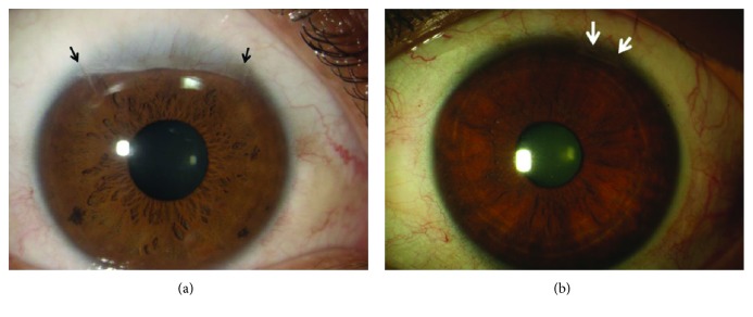
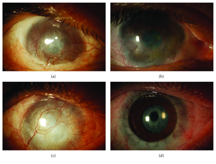
Limbal stem cell deficiency (LSCD) is a clinical condition characterized by damage of cornea limbal stem cells, which results in an impairment of corneal epithelium turnover and in an invasion of the cornea by the conjunctival epithelium. In these patients, the conjunctivalization of the cornea is associated with visual impairment and cornea transplantation has poor prognosis for recurrence of the conjunctivalization. Current treatments of LSCD are aimed at replacing the damaged corneal stem cells in order to restore a healthy corneal epithelium. The autotransplantation of limbal tissue from the healthy, fellow eye is effective in unilateral LSCD but leads to depauperation of the stem cell reservoir. In the last decades, novel techniques such as cultivated limbal epithelial transplantation (CLET) have been proposed in order to reduce the damage of the healthy fellow eye. Clinical and experimental evidence showed that CLET is effective in inducing long-term regeneration of a healthy corneal epithelium in patients with LSCD with a success rate of 70%-80%. Current limitations for the treatment of LSCD are represented by the lack of a marker able to unequivocally identify limbal stem cells and the treatment of total, bilateral LSCD which requires other sources of stem cells for ocular surface reconstruction.
Figures


Similar articles
-
Limbal stem cell transplantation: current perspectives.Clin Ophthalmol. 2016 Apr 1;10:593-602. doi: 10.2147/OPTH.S83676. eCollection 2016. Clin Ophthalmol. 2016. PMID: 27099468 Free PMC article. Review.
-
Cultivated limbal and oral mucosal epithelial transplantation.Semin Ophthalmol. 2012 May-Jul;27(3-4):80-93. doi: 10.3109/08820538.2012.680641. Semin Ophthalmol. 2012. PMID: 22784272
-
A Review of the Diagnosis and Treatment of Limbal Stem Cell Deficiency.Front Med (Lausanne). 2022 May 25;9:836009. doi: 10.3389/fmed.2022.836009. eCollection 2022. Front Med (Lausanne). 2022. PMID: 35692544 Free PMC article. Review.
-
Ocular surface reconstruction of Steven Johnson syndrome / toxic epidermal necrolysis affected eye - A case report.Heliyon. 2022 Dec 23;9(1):e12590. doi: 10.1016/j.heliyon.2022.e12590. eCollection 2023 Jan. Heliyon. 2022. PMID: 36820177 Free PMC article.
-
Simple limbal epithelial transplantation (SLET) in failed cultivated limbal epithelial transplantation (CLET) for unilateral chronic ocular burns.Br J Ophthalmol. 2018 Dec;102(12):1640-1645. doi: 10.1136/bjophthalmol-2017-311506. Epub 2018 Feb 16. Br J Ophthalmol. 2018. PMID: 29453224
Cited by
-
Corneal regeneration: insights in epithelial stem cell heterogeneity and dynamics.Curr Opin Genet Dev. 2022 Dec;77:101981. doi: 10.1016/j.gde.2022.101981. Epub 2022 Sep 6. Curr Opin Genet Dev. 2022. PMID: 36084496 Free PMC article. Review.
-
Subconjunctival Injection of Transdifferentiated Oral Mucosal Epithelial Cells for Limbal Stem Cell Deficiency in Rats.J Histochem Cytochem. 2021 Mar;69(3):177-190. doi: 10.1369/0022155420980071. Epub 2020 Dec 21. J Histochem Cytochem. 2021. PMID: 33345682 Free PMC article.
-
Matrix-Assisted Cell Transplantation for the Treatment of Limbal Stem Cell Deficiency in a Rabbit Model.Biomedicines. 2024 Jan 3;12(1):101. doi: 10.3390/biomedicines12010101. Biomedicines. 2024. PMID: 38255207 Free PMC article.
-
Priming human adipose-derived mesenchymal stem cells for corneal surface regeneration.J Cell Mol Med. 2021 Jun;25(11):5124-5137. doi: 10.1111/jcmm.16501. Epub 2021 May 5. J Cell Mol Med. 2021. PMID: 33951289 Free PMC article.
-
Therapeutic Strategies for Restoring Perturbed Corneal Epithelial Homeostasis in Limbal Stem Cell Deficiency: Current Trends and Future Directions.Cells. 2022 Oct 16;11(20):3247. doi: 10.3390/cells11203247. Cells. 2022. PMID: 36291115 Free PMC article. Review.
References
Publication types
LinkOut - more resources
Full Text Sources
