

Marginal Integrity of Cervical Restorations with Caries-Affected Dentinal Walls: Effect of Contamination with Hemostatic Agents
- PMID: 30405730
- PMCID: PMC6218462
Marginal Integrity of Cervical Restorations with Caries-Affected Dentinal Walls: Effect of Contamination with Hemostatic Agents
Abstract
Objectives: The aim of this study was to compare the microleakage in normal and caries-affected dentin (CAD) and to investigate the effect of three hemostatic agents on the microleakage of Class V composite resin restorations in CAD.
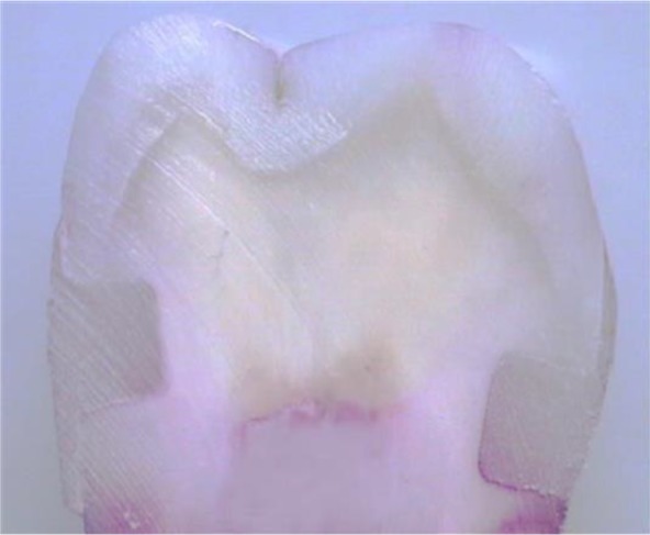
Materials and methods: Ninety-six Class V non-beveled cavities were prepared in 48 third molars at 1 mm below the cementoenamel junction (CEJ) in the cervical margin with the occlusogingival size of 2 mm, mesiodistal dimension of 3 mm, and a depth of 1.5 mm. Next, the teeth were divided into 8 groups (n=12): G1-4 included normal dentin (N) substrate, while G5-8 were exposed to mineralization/demineralization cycles to produce CAD substrate. Groups 1 and 5 were the controls. ViscoStat was used in groups 2 and 6, ViscoStat Clear was used in groups 3 and 7, while trichloroacetic acid (TCA) was used in groups 4 and 8. The cavities were restored with composite resin. The samples were sectioned after thermocycling and immersion in 2% fuchsin for 24 hours. The degree of dye penetration was evaluated under a stereomicroscope at 40× magnification. Data were evaluated using Kruskal-Wallis and Mann-U-Whitney tests in SPSS 15 software (α=0.05).
Results: Significant differences were recorded on the mean microleakage of different groups (P=0.047). There was a significant difference in the mean dentinal microleakage between N and CAD groups (P=0.014). The dentinal microleakage in group 3 was significantly higher than that in groups 4 to 8.
Conclusions: According to the results, CAD showed less microleakage in comparison with intact dentin. ViscoStat Clear caused a greater microleakage than did ViscoStat or TCA.
Keywords: Dental Leakage; Dentin-Bonding Agents; Hemostatics.
Figures
Similar articles
-
Sealing of adhesive systems in ferric sulfate-contaminated dentinal margins in class V composite resin restorations.J Dent Res Dent Clin Dent Prospects. 2016 Winter;10(1):17-22. doi: 10.15171/joddd.2016.003. Epub 2016 Mar 16. J Dent Res Dent Clin Dent Prospects. 2016. PMID: 27092210 Free PMC article.
-
Marginal integrity of low-shrinkage and methacrylate-based composite resins: Effect of three different hemostatic agents.J Clin Exp Dent. 2016 Apr 1;8(2):e178-83. doi: 10.4317/jced.52782. eCollection 2016 Apr. J Clin Exp Dent. 2016. PMID: 27034759 Free PMC article.
-
Comparison of Microleakage of Class V Cavities restored with the Embrace WetBond Class V Composite Resin and Conventional Opallis Composite Resin.J Contemp Dent Pract. 2017 Oct 1;18(10):867-873. doi: 10.5005/jp-journals-10024-2141. J Contemp Dent Pract. 2017. PMID: 28989122
-
[Comparative evaluation of marginal microleakage of three different resins in Class V composite restorations].Shanghai Kou Qiang Yi Xue. 2017 Jun;26(3):241-245. Shanghai Kou Qiang Yi Xue. 2017. PMID: 29098237 Chinese.
-
Gingival microleakage of class II bulk-fill composite resin restorations.Dent Med Probl. 2018 Oct-Dec;55(4):383-388. doi: 10.17219/dmp/99264. Dent Med Probl. 2018. PMID: 30648363
Cited by
-
Effect of contamination of bulk-fill flowable resin composite with different contaminants during packing on its surface microhardness and compressive strength: in vitro study.BMC Oral Health. 2022 Oct 17;22(1):446. doi: 10.1186/s12903-022-02495-6. BMC Oral Health. 2022. PMID: 36253744 Free PMC article.
References
-
- Siso HS, Kustarci A, Goktolga EG. Microleakage in resin composite restorations after antimicrobial pre-treatments: effect of KTP laser, chlorhexidine gluconate and Clearfil Protect Bond. Oper Dent. 2009. May-Jun;34(3):321–7. - PubMed
-
- Kimmes NS, Olson TL, Shaddy RS, Latta MA. Effect of ViscoStat and ViscoStat Plus on composite shear bond strength in the presence and absence of blood. J Adhes Dent. 2006. December;8(6):363–6. - PubMed
LinkOut - more resources
Full Text Sources
Miscellaneous