. 2018 Nov;16(5):6458-6464.
doi: 10.3892/ol.2018.9465. Epub 2018 Sep 20.

Lectin histochemical analysis of uterine natural killer cells in normal, hydatidiform molar and invasive molar pregnancy

Affiliations

Lectin histochemical analysis of uterine natural killer cells in normal, hydatidiform molar and invasive molar pregnancy

Ting Zhong et al. Oncol Lett. 2018 Nov.

Abstract

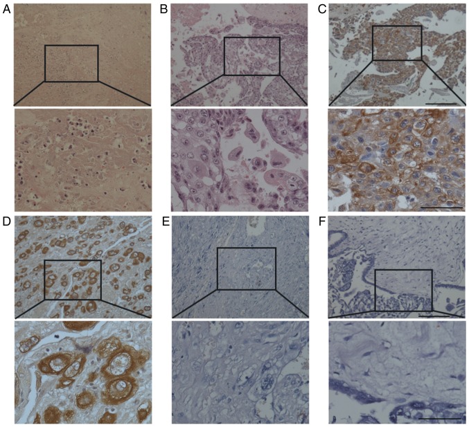
Uterine natural killer (uNK) cells have been hypothesized to serve a role in controlling trophoblast invasion and proliferation. The aim of the present study was to identify the distribution and number of uNK cells in normal pregnancy (NP), partial mole (PM), complete mole (CM) and invasive mole (IM). uNK cells were detected using dolichos biflorus agglutinin lectin immunohistochemistry in decidual and villous tissues from early NP (n=15), late NP (n=15), PM (n=22), CM (n=20) and IM (n=10). A scaled eye piece was used for cell counting to obtain semi-quantitative results. It was revealed that uNK cells were mainly located in the uterine deciduas of early NP. As pregnancy progressed, the number of decidual uNK cells significantly decreased. Decidual uNK cells of PM, CM and IM were located near blood vessel endothelial cells. No significant differences were detected with respect to the numbers of decidual uNK between early NP and PM. However, the number of decidual uNK cells was significantly reduced in CM and IM compared with early NP. The populations of decidual uNK cells were not significantly different between CM and IM. No uNK cells were detected in the villi of PM, CM or IM. The decrease of decidual uNK cells in late NP, CP and IM, compared with early NP, suggested that uNK cells served an important role in controlling trophoblast invasion and proliferation.

Keywords: decidua; dolichos biflorus agglutinin lectin; gestational trophoblastic disease; uterine natural killer cells.


Figures

Figure 1.
Figure 1.
Distribution of uNK cells at the maternal-fetal interface in the first trimester normal pregnancy. Trophoblast cell-identifying cytokeratin staining of (A) villous and (B) decidual tissue. (C) Mouse uterus tissue at day 8 of normal pregnancy, used as a positive control. Brown-yellow staining indicates positive DBA lectin staining of uNK cells. (D) Photomicrograph of DBA lectin-stained villous sections from the first trimester of normal pregnancy. (E-F) Photomicrographs of DBA lectin-stained decidual sections from the first trimester of normal pregnancy indicating (E) large mature uNK cells and (F) smaller immature NK cells. Each tissue is represented at magnification, ×200 (above) and ×400 (below). Inset of F (below) shows immunostaining of a negative control-stained section (primary antibody was replaced by 0.1 M N-acetyl-D-galactosamine). uNK, uterine natural killer; DBA, dolichos biflorus agglutinin. Scale bar, 50 µm.
Figure 2.
Figure 2.
Distribution of uNK cells at the normal maternal-fetal interface in term pregnancy. (A) Photomicrograph of DBA lectin-stained decidual tissue section of a normal term pregnancy. (B) Photomicrograph of DBA lectin-stained villous section from normal term pregnancy. Each tissue is represented at magnification, ×200 (above) and ×400 (below). uNK, uterine natural killer; DBA, dolichos biflorus agglutinin. Scale bar, 50 µm.
Figure 3.
Figure 3.
Distribution of uNK cells in partial hydatidiform mole. Hematoxylin and eosin staining of (A) decidual and (B) villous tissue of partial hydatidiform mole. Cytokeratin staining of (C) decidual and (D) villous tissue of hydatidiform mole to identify trophoblast cells. Brown staining indicates cytokeratin protein expression. Photomicrographs of DBA lectin staining of (E) decidual and (F) villous tissue of partial hydatidiform mole. Each tissue is represented at magnification, ×200 (above) and ×400 (below). uNK, uterine natural killer; DBA, dolichos biflorus agglutinin. Scale bar, 50 µm.
Figure 4.
Figure 4.
Distribution of uNK cells in complete hydatidiform mole. Hematoxylin and eosin staining of (A) decidual and (B) villous tissue of complete hydatidiform mole. Cytokeratin staining of (C) decidual and (D) villous tissue to identify trophoblast cell. Brown staining indicates cytokeratin protein expression. Photomicrographs of DBA lectin staining of (E) decidual and (F) villous tissue of complete hydatidiform mole. Each tissue is represented at magnification, ×200 (above) and ×400 (below). uNK, uterine natural killer; DBA, dolichos biflorus agglutinin. Scale bar, 50 µm.
Figure 5.
Figure 5.
Distribution of uNK cells in invasive hydatidiform mole. Hematoxylin and eosin staining of (A) decidual and (B) and villous tissue of invasive hydatidiform mole. Cytokeratin staining of (C) decidual and (D) villous tissue to identify trophoblast cell. Brown staining indicates cytokeratin protein expression Photomicrographs of DBA lectin staining of (E) decidual and (F) villous tissue of complete hydatidiform mole. Each tissue is represented at magnification, ×200 (above) and ×400 (below). uNK, uterine natural killer; DBA, dolichos biflorus agglutinin. Scale bar, 50 µm.

References

    1. Cheung AN. Pathology of gestational trophoblastic diseases. Best Pract Res Clin Obstet Gynaecol. 2003;17:849–868. doi: 10.1016/S1521-6934(03)00094-4. - DOI - PubMed
    1. Monchek R, Wiedaseck S. Gestational trophoblastic disease: An overview. J Midwifery Womens Health. 2012;57:255–259. doi: 10.1111/j.1542-2011.2012.00177.x. - DOI - PubMed
    1. Seckl MJ, Sebire NJ, Berkowitz RS. Gestational trophoblastic disease. Lancet. 2010;376:717–729. doi: 10.1016/S0140-6736(10)60280-2. - DOI - PubMed
    1. Tse KY, Chan KKL, Tam KF, Ngan HYS. Gestational trophoblastic disease. Obstet Gynaecol Reprod Med. 2009;19:89–97. doi: 10.1016/j.ogrm.2008.12.002. - DOI
    1. Bagshawe KD, Lawler SD, Paradinas FJ, Dent J, Brown P, Boxer GM. Gestational trophoblastic tumours following initial diagnosis of partial hydatidiform mole. Lancet. 1990;335:1074–1076. doi: 10.1016/0140-6736(90)92641-T. - DOI - PubMed