. 2018 Nov;16(5):6582-6588.
doi: 10.3892/ol.2018.9462. Epub 2018 Sep 19.

Invasive bronchial fibroblasts derived from asthmatic patients activate lung cancer A549 cells in vitro

Affiliations

Invasive bronchial fibroblasts derived from asthmatic patients activate lung cancer A549 cells in vitro

Damian Ryszawy et al. Oncol Lett. 2018 Nov.

Abstract

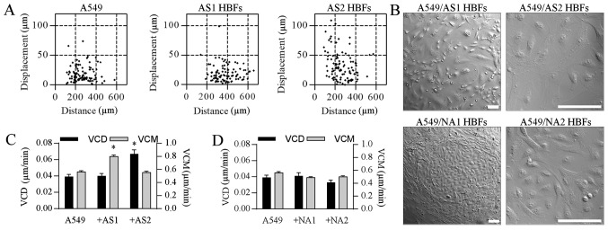
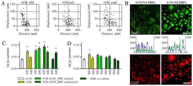
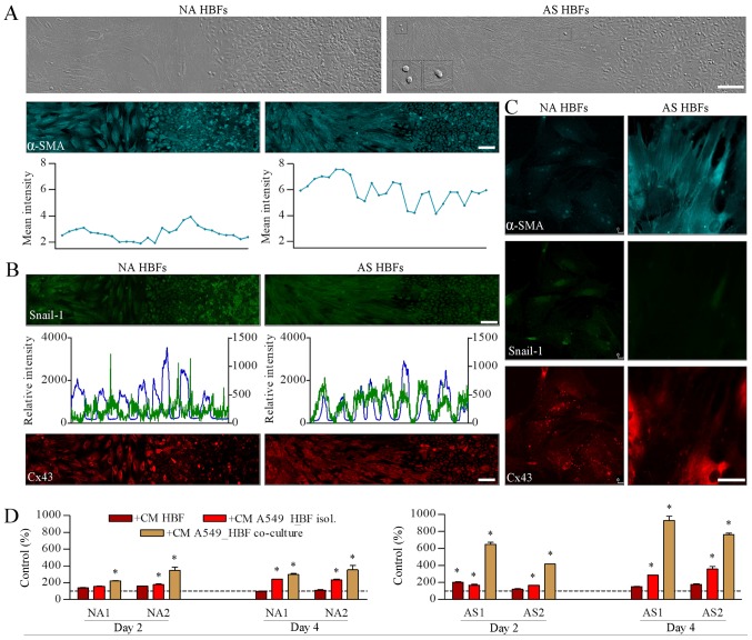
Epidemiological data suggests that there are functional links between bronchial asthma and lung carcinogenesis. Bronchial fibroblasts serve a prominent role in the asthmatic process; however, their involvement in lung cancer progression remains unaddressed. To estimate the effect of the asthmatic microenvironment on the invasiveness of lung cancer cells, the present study compared the behavior of human non-small cell lung cancer A549 cells exposed to the signals from human bronchial fibroblasts (HBFs) derived from non-asthmatic donors (NA HBFs) and from asthmatic patients (AS HBFs). NA HBFs did not significantly affect A549 motility, whereas AS HBFs and the media conditioned with AS HBF/A549 co-cultures increased Snail-1/connexin43 expression and motility of A549 cells. In contrast to NA HBFs, which formed A549-impenetrable lateral barriers, α-SMA+ AS HBFs actively infiltrated A549 monolayers and secreted chemotactic factors that arrested A549 cells within AS HBF/A549 contact zone. However, small sub-populations of A549 cells could release from this arrest and colonize distant regions of AS HBF monolayers. These data indicated that the interactions between lung cancer cells and HBFs in asthmatic bronchi may facilitate the colonization of lung tumors by fibroblasts. It further stabilizes the tumor microenvironment and potentially facilitates collective colonization of novel bronchial loci by cancer cells. Potential mechanistic links between the asthmatic process and lung cancer progression suggest that bronchial asthma should be included in the list of potential prognostic markers for lung cancer therapy.

Keywords: CAFs; bronchial asthma; intercellular communication; invasion; lung cancer.


Figures

Figure 1.
Figure 1.
HBFs stimulate the invasive behavior of A549 cells. (A) A549 cells and AS HBFs were seeded at a density of 104 and 103 cells/cm2, respectively, and were incubated for 48 h in Dulbecco's modified Eagle's medium supplemented with a 10% fetal calf serum. The motility of A549 cells was estimated with time-lapse videomicroscopy and (C) its parameters were quantified in comparison to control A549 cells. (B) A549/HBF co-cultures were established as in (A) and cell morphology was visualized with NIC microscopy. Scale bars, 100 µm. (D) The motility of A549 cells in co-cultures with NA HBFs estimated by time-lapse videomicroscopy as in (A). At least 50 cell trajectories were drawn for each condition and presented in correlative plots. Dot-plots and column charts present movement parameters at the single cell and population level, respectively. Data are representative of 3 independent experiments. *P<0.05 vs. A549. Data are presented as the mean ± standard error of the mean. Note that the disruption and penetration of A549 cell monolayers by AS HBFs is correlated with the induction of A549 motility. HBFs, human bronchial fibroblasts; AS, asthmatic donors; NA, non-asthmatic donors; VCM, velocity of cell motility; VCD, velocity of cell displacement; NIC, Nomarski Interference Contrast.
Figure 2.
Figure 2.
AS HBFs induce the motility of A549 cells via contact-modulated paracrine signaling. (A) A549 cells were cultivated in the media conditioned by AS2 HBFs (left), ‘separated’ (middle) and ‘confronted’ AS HBF/A549 co-cultures (1:1 v/v with fresh medium) for 48 h and (C) the parameters of their motility were analyzed by time-lapse videomicroscopy in comparison to A549 motility in control conditions and in ‘open’ AS HBF/A549 co-cultures. (B) A549 cells were cultivated in the media conditioned by ‘separated’ co-cultures of A549 with AS and NA HBFs (1:1 v/v with fresh medium) for 48 h. Intracellular localization of Snail-1/Cx43 and co-localization of Snail-1/DNA was visualized with immunofluorescence and cytofluorimetry, respectively (left axes/blue line: DNA; right axes/green line: Snail-1). Scale bar, 50 µm; magnification, ×400. (D) The motility of A549 cells in the presence of the media conditioned by NA HBFs (left), ‘separated’ (middle) and ‘confronted’ NA HBF/A549 co-cultures (1:1 v/v with fresh medium) was analyzed as in (A). Data are presented as the mean ± standard error of the mean of 3 independent experiments. *P<0.05 vs. A549. Note the mobilization of A549 in the presence of AS HBF/A549-conditioned media, which was correlated with Snail-1/Cx43 activation. HBFs, human bronchial fibroblasts; AS, asthmatic donors; NA, non-asthmatic donors; VCD, velocity of cell displacement; Cx43, connexin43; CM, conditional media.
Figure 3.
Figure 3.
AS HBFs selectively modulate the invasion of A549 cells. (A) A549 cells were grown to confluence with NA HBFs (left-hand panel) or AS HBFs (right-hand panel) in 2-well silicone inserts and co-incubated for 48 h following diaphragm removal prior to fixation and immunostaining for α-SMA. Expression of α-SMA was visualized by immunofluorescence and cytofluorimetry, respectively. Scale bar, 100 µm; magnification, ×200. Inserts present A549 cells within the HBF monolayer. (B) Cells were cultivated as in (A) and intracellular localization of Snail-1/Cx43 and co-localization of Snail-1/DNA were visualized with immunofluorescence and cytofluorimetry, respectively (left axes: DNA; right axes: Snail-1). Photomicrographs in (A) and (B) present panoramic till scans of the interfaces between HBF and A549 monolayers obtained by the composition of two rows of succeeding images (7 pictures in a row). Scale bars, 100 µm. (C) NA HBFs and AS HBFs were cultivated in the presence of the media conditioned by ‘separated’ NA HBF/A549 and AS HBF/A549 co-cultures, respectively, for 48 h and immunostained against α-SMA, Snail-1 and Cx43. Scale bars, 50 µm; magnification, ×630. (D) A549 cells were seeded onto microporous membrane, placed in the wells filled with the media conditioned by A549/HBF co-cultures, and allowed to transmigrate for 48 h. The number of the transmigrated cells were counted following 24 h. Data are presented as the mean ± standard error of the mean of 3 independent experiments. *P<0.05 vs. A549 parameters in control medium. Note the infiltration of AS HBFs into A549 monolayers, accompanied by the chemotactic activity of the media from ‘open’ AS HBF/A549 co-cultures and by the presence of scattered A549 within AS HBF continua. HBFs, human bronchial fibroblasts; AS, asthmatic donors; NA, non-asthmatic donors; α-SMA, α-smooth muscle actin; Cx43, connexin43; CM, conditional media.

Similar articles

Cited by

References

    1. Anandan C, Nurmatov U, van Schayck OC, Sheikh A. Is the prevalence of asthma declining? Systematic review ofepidemiological studies. Allergy. 2010;65:152–167. doi: 10.1111/j.1398-9995.2009.02244.x. - DOI - PubMed
    1. Al Muhsen S, Johnson JR, Hamid Q. Remodeling in asthma. J Allergy Clin Immunol. 2011;128:451–462. doi: 10.1016/j.jaci.2011.04.047. - DOI - PubMed
    1. Halwani R, Al Muhsen S, Al Jahdali H, Hamid Q. Role of transforming growth factor-β in airway remodeling in asthma. Am J Respir Cell Mol Biol. 2011;44:127–133. doi: 10.1165/rcmb.2010-0027TR. - DOI - PubMed
    1. Ingram JL, Huggins MJ, Church TD, Li Y, Francisco DC, Degan S, Firszt R, Beaver DM, Lugogo NL, Wang Y, et al. Airway fibroblasts in asthma manifest an invasive phenotype. Am J Respir Crit Care Med. 2011;183:1625–1632. doi: 10.1164/rccm.201009-1452OC. - DOI - PMC - PubMed
    1. Darby IA, Laverdet B, Bonté F, Desmoulière A. Fibroblasts and myofibroblasts in wound healing. Clin Cosmet Investig Dermatol. 2014;7:301–311. - PMC - PubMed