

Adipsin, MIP-1b, and IL-8 as CSF Biomarker Panels for ALS Diagnosis
- PMID: 30405855
- PMCID: PMC6199888
- DOI: 10.1155/2018/3023826
Adipsin, MIP-1b, and IL-8 as CSF Biomarker Panels for ALS Diagnosis
Abstract
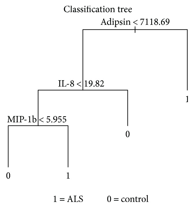
Amyotrophic lateral sclerosis (ALS) is an aggressive neurodegenerative disorder that selectively attacks motor neurons in the brain and spinal cord. Despite important advances in the knowledge of the etiology and progression of the disease, there are still no solid grounds in which a clinician could make an early objective and reliable diagnosis from which patients could benefit. Diagnosis is difficult and basically made by clinical rating scales (ALSRs and El Escorial). The possible finding of biomarkers to aid in the early diagnosis and rate of disease progression could serve for future innovative therapeutic approaches. Recently, it has been suggested that ALS has an important immune component that could represent either the cause or the consequence of the disease. In this report, we analyzed 19 different cytokines and growth factors in the cerebrospinal fluid of 77 ALS patients and 13 controls by decision tree and PanelomiX program. Results showed an increase of Adipsin, MIP-1b, and IL-6, associated with a decrease of IL-8 thresholds, related with ALS patients. This biomarker panel analysis could represent an important aid for diagnosis of ALS alongside the clinical and neurophysiological criteria.
Figures


References
-
- Tanaka M., Kikuchi H., Ishizu T., et al. Intrathecal upregulation of granulocyte colony stimulating factor and its neuroprotective actions on motor neurons in amyotrophic lateral sclerosis. Journal of Neuropathology & Experimental Neurology. 2006;65(8):816–825. doi: 10.1097/01.jnen.0000232025.84238.e1. - DOI - PubMed
MeSH terms
Substances
LinkOut - more resources
Full Text Sources
Medical
Miscellaneous
